Click on IMAGE to enlarge photos. Click on BLUE link to read articles.
AUSTIN FLINT: THE "AMERICAN LAENNEC"
Austin Flint, Sr., M.D., who was recognized as an execptional clinician and teacher, prolific writer, visionary thinker and the forerunner of modern cardiology, was regarded as the "American Laennec" (Dr. Samuel Gross). Austin Flint was a graduate of Havard Medical School where he was a pupil of James Jackson, who was an early advocate of the use of the stethoscope for ausculattion. Dr. Flint was one of the founders of the Buffalo Medical College and Bellevue Hospital Medical College, also taught in Chicago, Louisville and New Orleans before settling in New York in 1861, serving as professor of both the Long Island College Hopsial (now SUNY Downstate Medical College) and and Bellevue Hospital Medical College (now NYU School of Medicine). He was elected president of the New York Academy of Medicine in 1872 and the American Medical Association in 1884. Dr. Flint continued to evolve the art of physical diagnosis and especially percussion and auscultation initiated earlier by Leopold Auenbrugger and Rene Laennec, respectively. His texts on diseases of the heart, respiratory system and manual on percussuoin and auscultation are considered classics. By the time of his death on March 13, 1886, he had a national and internatioal reputation as an esteemed medical professional.
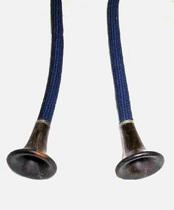
In 1852, George P. Cammann, M.D. desiged a stethoscope that used both ears. This new "binaural" instrument was touted to be the preferred method of auscultation. Also in 1852, Dr. Austin Flint published his prize essay On Variations in Pitch in Percussion and Respiratory Sounds and their Application to Physical Dignosis establishing his expertise in ausculataion of the chest. At first, Dr. Flint was concerned that the transmission of sounds by the binaural stethoscope would be attenuated and that its advantages were not appreciated without considerable practice. In his text on Physical Exploration and Diagnosis of Diseases affecting the Respiratory Organs published in 1856 he stated "In making trial of this instrument, I have found it more difficult to institue comparisons as regards quality and pitch of sound with the ear alone, or the ordinary stethoscope." Ten years later in the second edition of his text publisheed in 1866, 3 years after Dr. Cammann's death, he corrected his opnion stating that "the objection on the score of the alteration of the pitch and quality of sounds I have long since found to be without foundation, and I am sure that this instrument will supplant all wooden stethosopes as soon as it is fully appreciated."
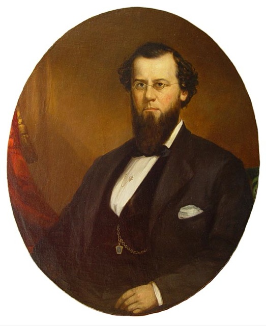
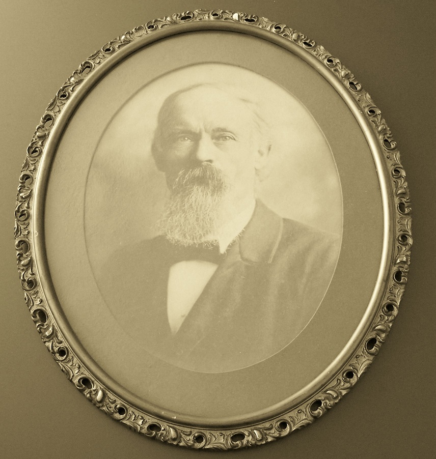
Oil portrait of
Austin Flint, Sr., M.D. shown on the left was a family portrait by the New York painter John L. Harding,
Sr. and is signed and dated 1865 by the artist. Dr. Flint is wearing a
pin of the Freemasons on his vest and Victory of the Cross on his shirt.
The Flint family donated the portrait to the New York Academy of
Medicine in 1900 and was displayed in their Museum Room. The New York
portraitist George
R.
Boynton was
commissioned by
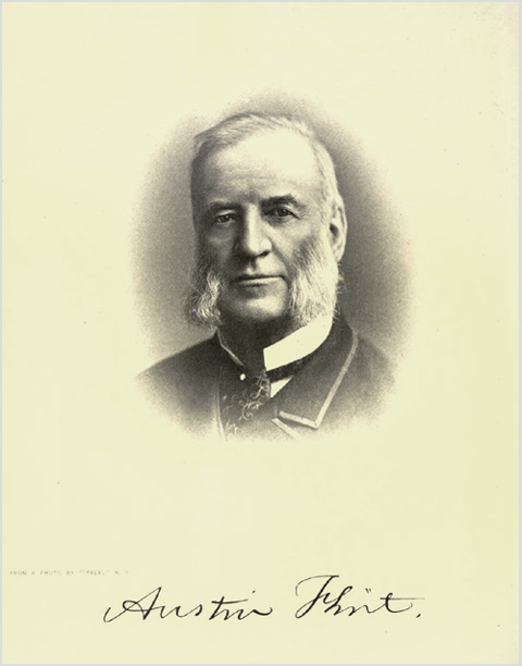
the New York Academy of Medicine to paint a posthumous portrait
of Dr. Flint shown in the center photo. This very portrait was
preseted
to the Academy on January 17, 1901 by Dr. J.E. Janeway on behalf of
himself and 14 other fellows of the Academy who were the donors for the
portrait. This posthumous oil portrait
was signed by Boynton and based on the last known photograph of Dr.
Flint by the New
York portraitist photographer Benjamin J. Falk, as
shown in The Medicine of the Future publication of Dr. Flint's intended
address
to the British Medical Association in 1886, delivered by Dr. Flint's
son, Austin Flint, Jr., on April 24, 1886 after his father's death.
Note that the paisley patterned ascot and stickpin in the painting are
the same as in the photopgraph. The portrait was displayed in the
President's Gallery at the New York Academy of Medicine. John Harding,
George Boynton and
Benjamin Falk were all leading New York artists that were well known
for their portraits
and especially their use of lighting and positioning of the subject.
( Photos courtesy of Alex Peck )
Actually, the idea for a binaural
stethoscope was first introduced in 1829, just ten years after the
publication of Laennec's text illustrated his original instrument. The
idea belonged to Nicholas Comins, who devised a stethoscope that he
described as "a bent tube" that had several hinges, allowing the
physician to not have to assume uncomfortable positions during the
examination. He offered the suggestion of making his instrument
binaural, but there are only sketches of his instrument.
Dr.
C.J.B. Willaims is said to have constructed a binaural stethoscope
using two bent pipes (since rubber was not yet available) attached
to a wood chest piece around 1840. Arthur Leared presented a model
of a "double" stethoscope made of gutta-percha at the Great Exhibition
in London in 1851. The first commertcially available binaural
stethoscope was made by Dr. Nathan B. Marsh of Cincinnati,
patented in 1851. His model was made of india rubber with a long
stem to which a flaring bell made of wood could be attached. However,
it proved cumbersome, very fragile and quickly faded.
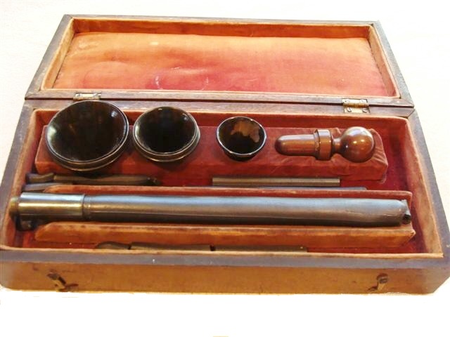

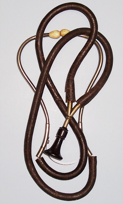
The
original Marsh binaural stethoscope, circa 1851. On the left the
stethoscope is in its original wood box. On the right is the assembled
stethoscope.
(photos courtesy of the Marsh family,
Smithsonian museum)
GEORGE CAMMANN: THE BINAURAL STETHOSCOPE
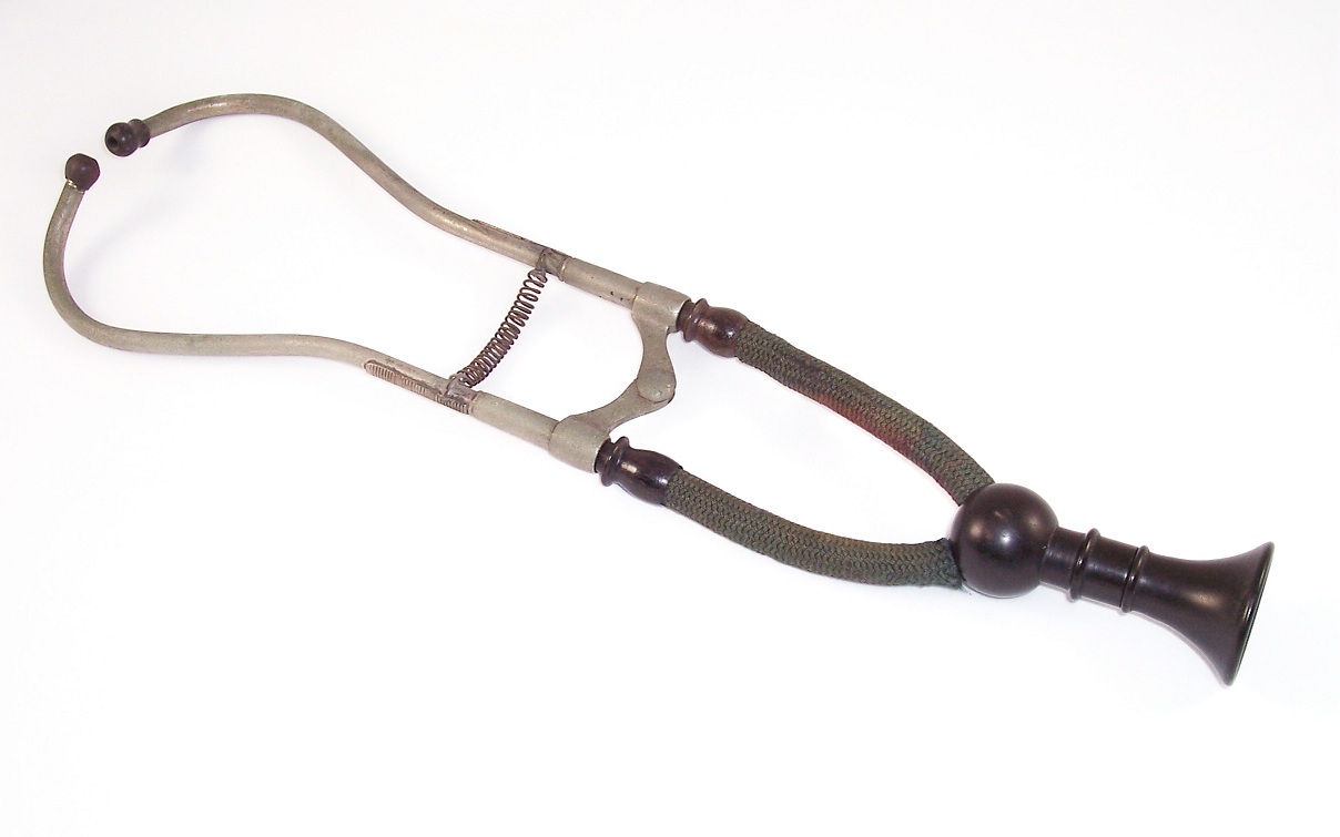
In
1852, Dr. George Cammann of New York produced the first recognized
usable binaural stethoscope. He was working as a physician at the
Northern Dispensary in New York City and had seen Marsh's model. He
also had a model of a soft metal, multiple-tubed stethoscope made by H.
Landouzy in 1841, which was designed for two people to listen at the
same time. And Charles J. B. Williams claims to have made a binaural
stethoscope with lead tubes in 1843. Cammann did not claim to have the
original idea for a binaural stethoscope, only to have developed a
practical instrument that could be used in clinical practice. Interestingly,
he never
patented the stethoscope believing it should be freely available to
physicians. The stethoscope was named Cammann's Stethoscope by the
manufacturer of the original instrument, George Tiemann. Cammann's
model was made with ivory earpieces connected to metal tubes of German
silver that were
held together by a simple hinge joint, and tension was applied by way
of an elastic band. Attached to these were two tubes covered by wound
silk. These converged into a hollow ball designed to amplify the sound,
and attached to the ball was a conical shaped, bell chest piece.


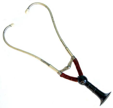
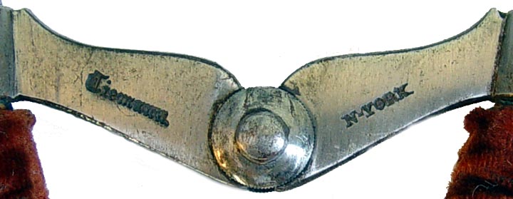

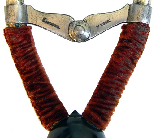
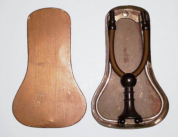
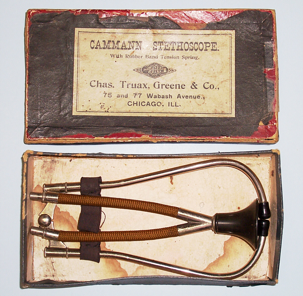
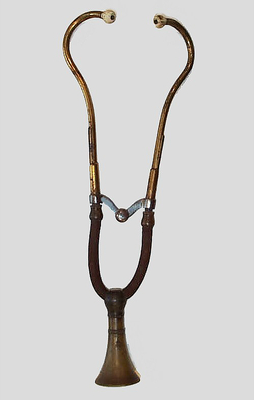
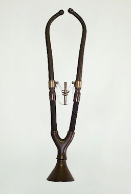
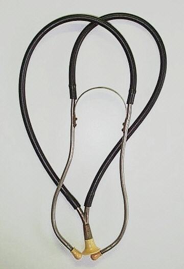
The earliest known original model Cammann binaural
stethoscope made by George Tiemann, circa 1852. This is an exceptionally
rare and important early 1850s initial model Cammann binaural
stethoscope with the front of the yoke delicately hand-engraved Dr.
Cammann's / Stethoscope. Tiemann / N. YORK is stamped on
the reverse side. Note the velvet sleeves covering the
very short tubes. The metal ear tubes are made of German
silver, large and flaring chestpiece was
turned from ebony wood, and the earpieces are ivory. This stethoscope
was obtained from the Intenational Museum of Surgical Science,
Chicago, Il, when it reorganized its science collection and is
the same instrument pictured in American Surgical Instruments, by James M. Edmonson , p. 106, figs. 138
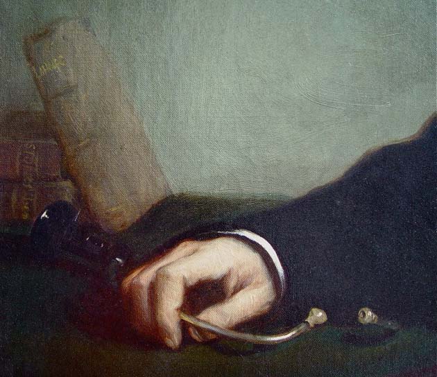
and 139. It is the stethoscope shown in the above detail of Dr.
Cammann's portait.
(Photos courtsey Alex Peck)
Prior
to 1855 George Tiemann marked his medical instruments as Tiemann, after
1855 used the mark G. Tiemann & Co. and later used the mark Tiemann
& Co. The markings on the models shown below help date these
stethoscopes and are consistent with the introduction of the Cammann
stethoscope in 1852.


![]()
![]()
![]()
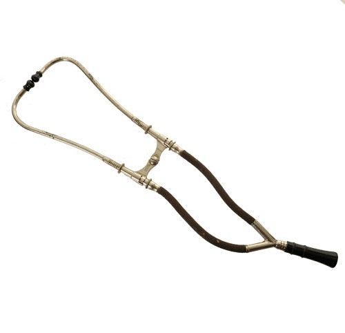
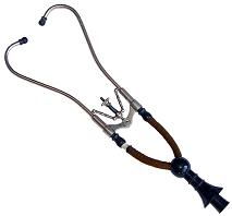
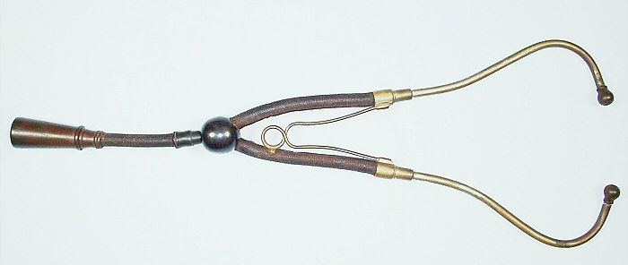
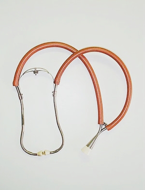
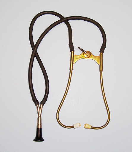
To the left is an original Cammann stethoscope, circa 1852. The yoke of the hinge joint is hand engraved Dr. Cammann's Stethoscope and the reverse is stamped Tiemann N[ew]. York. This initial model had a large and flaring ebony chestpiece and ivory earpieces. The flexible woven tubes were covered with velvet and were very short as compared to later models. This stethoscope and the one above are the only two hand engraved Cammann stethoscopes known to exist.
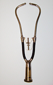
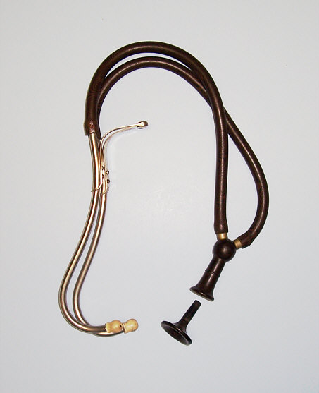
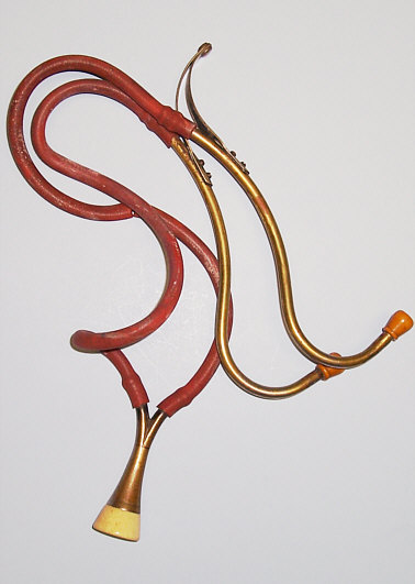
The Cammann stethoscope shown in the middle photo dates to 1855. The left side of the yoke of the hinge joint is stamped Dr. Cammann's Stethoscope, but is not hand engraved. The right side is stamped G. Tiemann & Co. Note that the wood bell does not have as large a flare and the velvet covered tubes are a little longer. This is the only example of this Cammann stethoscope known to exist.
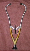
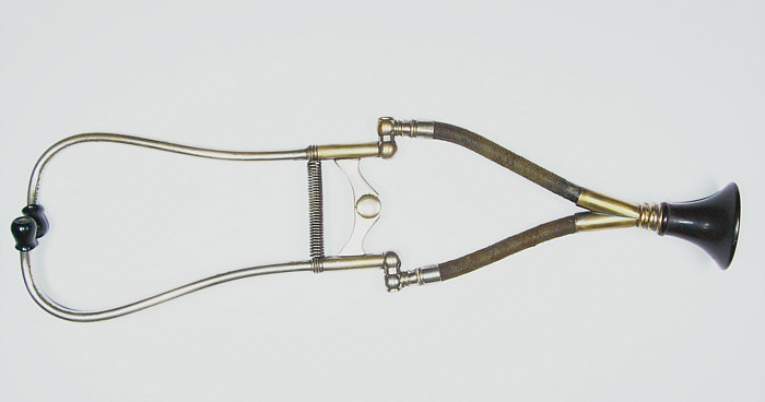
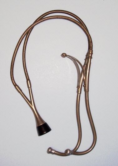
On the right is Cammann stethoscope, circa 1860. The left side of the yoke is marked Dr. Cammann's Stethoscope but is not hand engraved. The right side is marked Tiemann & Co. Note that the flexible tubes are longer, not covered in velvet and are attached by gutta percha connectors to the binaural ear tubes. The two parts of the stethoscope could be detached for carrying and reattached for auscultation.


On the left is the only known
carte-de-viste (cdv) albumen print photograph of Dr. Cammann by Matthew
B. Brady & Studio
in New York City, circa 1860, which appears to have been taken at the
same sitting as the salt print photo mentioned above.
Note the Brady backmark on the verso as well as the period ink
signature of G. P. Cammann, M.D. below the backmark. Dr. Cammann
developed the binaural stethoscope in 1852 while
working at the
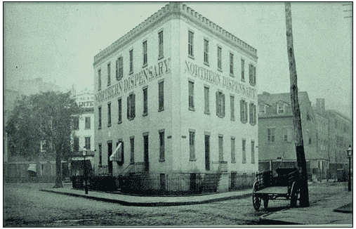
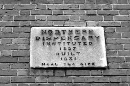
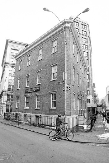
Northern Dispensary. The Northern Dispensary was instituted in 1827 and
built in 1831 to
provide medical care to the "sick and worthy poor" and was open
continuously from 1831
until 1989. The Dispensary is the only building in New York
City that has two sides on one street (Waverly Place) and one side on
two streets (Christopher and Grove Streets). The three sided building
occupies the triangle formed by those streets. Shown above are the
photos of the Northern Dispensary in 1855 after addition of a third
floor to the 1831 building and in 2006 looking the same as it did in
1855, but now closed and empty for 25 years because the original deed
requires that the building only be used for aiding the sick and worthy
poor. Notice the
young boy pausing on his bike, while trying to decide which fork in the
Waverly Place road to take, which splits into a left and right street
at the tip of the Northern Dispensary triangle. Dr. Cammann was
appointed to the Dispensary in 1832 as an Attending Physician and
worked there for 28 years until he resigned in 1859 and then contiunued
to serve as a Consulting Physician until his death in 1863. Shown
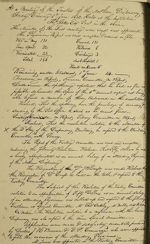
above are photos of pages from the June 7, 1832 and October 7,
1859 minutes of the Proceedings of the Trustees of the Northern
Dispensary appointing Dr. Cammann to the Dispensary and thanking Dr.
Cammann for his faithful service to the Dispensary, respectively. He
was also a member of the St. James Episcopal Church, Fordham and one of
the church's four Royal Bavarian Stained Glass Windows
portraying Saints John and Peter healing of a man by the
Jerusalem Temple's Beautiful Gate, installed when the church was
built on its present site in 1864-65, was in memory of Dr. Cammann for
his humanitarianism. The window dedication reads "In Memory of Geo. P.
Camann M.D. dec. Feb. 14. 1863."
(Nothern Dispensary minutes
courtesy of the New York University Archives, Northern Dispensary
photo by Jefferson Siegel and stained glass window photo courtesy of
St. James Episcopal Church, Fordham).


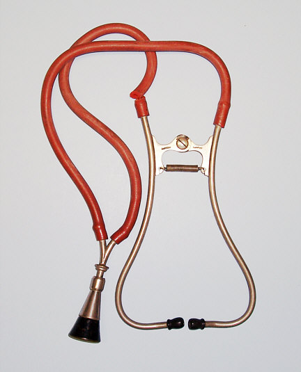
To
the far left is Cammann stethoscope with elastic band, which was the
original tension mechanism designed by Dr. Cammann, W.F. Ford & Co,
circa 1870. Next is Cammann stethoscope with spring tension mechanism,
circa 1880 and a Cammann stethoscope with Ford's patented wire
spring tension mechanism to hold ear peices together, Hazard,
Harzard & Co, circa 1890. On the far right is a Cammann stethoscope
with screw tension mechanism, Sharp & Smith, circa 1880.
Cammann stethosope in its original tin
case, circa 1870.
The
Cammann stethoscope usually came in a carrying case, although most of
these were lost over time. The cases are more difficult to find than
the actual stethoscopes. Two examples of such cases
are shown below.

Cammann stethoscope, circa 1880, marked Dr. Cammann Stethoscope, Tiemann Co. The tooled leather case is marked Dr. John Hatton (1838-1898) who graduated the Iowa University Medical College in 1870 and practiced in Des Moines.
As the Cammann stethoscope evolved, diffirent materials were used for the metal tubes and bell. Originally metal tubes were made of German silver and chest pieces of ebony wood. Later models of the Cammann stethoscope became simplier in the shape of the bell and metal attachment of the flexible tubes to the metal tubes.

Turn of the century Cammann stethoscope
with small ebony bell marked Weiss, London circa 1901.
As
was the case with Laennec's model, Cammann's was not embraced
completely for quite some time. It was not until Austin Flint (who had
previously spoken against the binaural in 1856) endorsed it in 1866
that it became widely used. During the later half of the 19th century,
well educated physicians used advances in medical technology to aid
their ability to diagnose diseases in their patients. The stethoscope
rapidly became the main symbol of the highly skilled physician.

A
photo dated May 1889 of a physician in his office with a medical text
on his lap, surrounded by medical equipment including a Bauch &
Lomb microscope and Cammann stethoscope on his desk. The use of the
microscope and stethoscope by physicians represents the advances in
technology applied to the practice of medicine in the nineteenth
century.
THE HUSE COLLECTION
The
Harvard Medical School (HMS) was founded in 1782 as the
Massachusettes Medical College in Cambridge. John Warren,
Professor of Anatomy and Surgery, and one of the three original medical
college faculty, was instrumetal in moving the school to Boston.
As there was no hospital in Boston at that time, teaching medical
students was devoid of clinical experience. In 1811, his son, John
Collins Warren, was a leader in establishing the
Massachusetts General Hospital (MGH), the third oldest hospital in the
United States. Like most hospitals founded in the 19th
century, MGH was intended to care for the poor. In 1869, a
progressive curriculum was instituted at the school,
new
departments of basic and clinical sciences were established, a
three-year degree program was introduced, and the apprenticeship system
was eliminated. Harvard Medical School became a professional school of
Harvard University, setting the United States standard for the
organization of medical education within a university.


Pictured
above in the middle is the Bulfinch Buiding of MGH, circa 1880, with
its
famous ether dome on top of the buiding. Ether was first used for
anesthesia during surgery at the MGH in 1846. The dome shaped and
glass ceiling of the surgical theater not only enabled surgeons to
operate with natural daylight but also to remain awake while performing
surgery on their sleeping patients as the fumes from ether rose to the
top of the domed amphitheater, as shown in the current photo of the
ether dome on the left. HMS moved off the Cambridge campus
to Boston and was located adjacent to MGH from 1846 through 1883. The
HMS is pictured in the foreground in the photo on right with the
adjacent MGH in the background, circa 1875. In 1886, Reginald H.
Fitz, Visiting Physician at MGH and the Shattuck Professor of
Anatomical Pathology at HMS, published his seminal paper on
"Perforating Inflammation of the Vermiform Appendix; with special
reference to its early diagnosis and treatment". For the first time the
term appendicitis was used to describe the most inflammatory disease of
the right lower quadrant of the abdomen which was caused by
inflammation of the appendix and that treatmant was early surgical
removal of the inflammed appendix.
The
unique medical collection displayed below is from Dr. Charles
Archelaus Huse's medical office circa 1884 in Worcester,
Massachusettes. The collection includes Dr. Huse's Cammann stethoscope,
surgical instrument kit, urethral silver catheters, patient records,
patient receipts, letter from the Dudley & Sheppard instrument
company dated 1877 where Dr. Huse purchased his medical
instruments, medical student ticket to Harvard Medical School
dated 1879-80, and sign from his office in Worcester. Dr.
Huse was born on August 7, 1855 in Worcester, graduated Brown
University in 1878 and Harvard Medical School in 1881. After
graduation from Harvard, he served as the Boston Board of
Health Assistant Port Physician for 14 months stationed at Deer
Island. In the summer of 1882, he moved back to his home town of
Worcester and established his practice of medicine and surgery. He
was a member of the Massachusetts Medical Society and the Worcester
Society for Medical Improvement from 1882-1884. Dr. Huse died
prematurely at age 29 from "peritonitis" after a nine day illness
on July 3, 1884, just two years prior to the publication of the classic
paper by his former Harvard Professor, Dr. Fitz,
identifying the cause of most cases of peritionitis
as the rupture of an inflammed appendix, which required
surgical resection as the definitve treatment. The
Huse antique medical collection represents a truly
unique look at this promising young physician and the practice of
medicine in the late 19th century.

The
middle photo shows Dr. Huse's outside office sign from 136 Austin
Street, Worcester, Massachusettes, circa 1884, along with a ticket
for C. A. Huse to attend Harvard Medical School in 1879-80. On the
left is a potrait cdv photo of Charles A. Huse as a student at Brown
University circa 1878 and on the right is a cabinet photo of him with
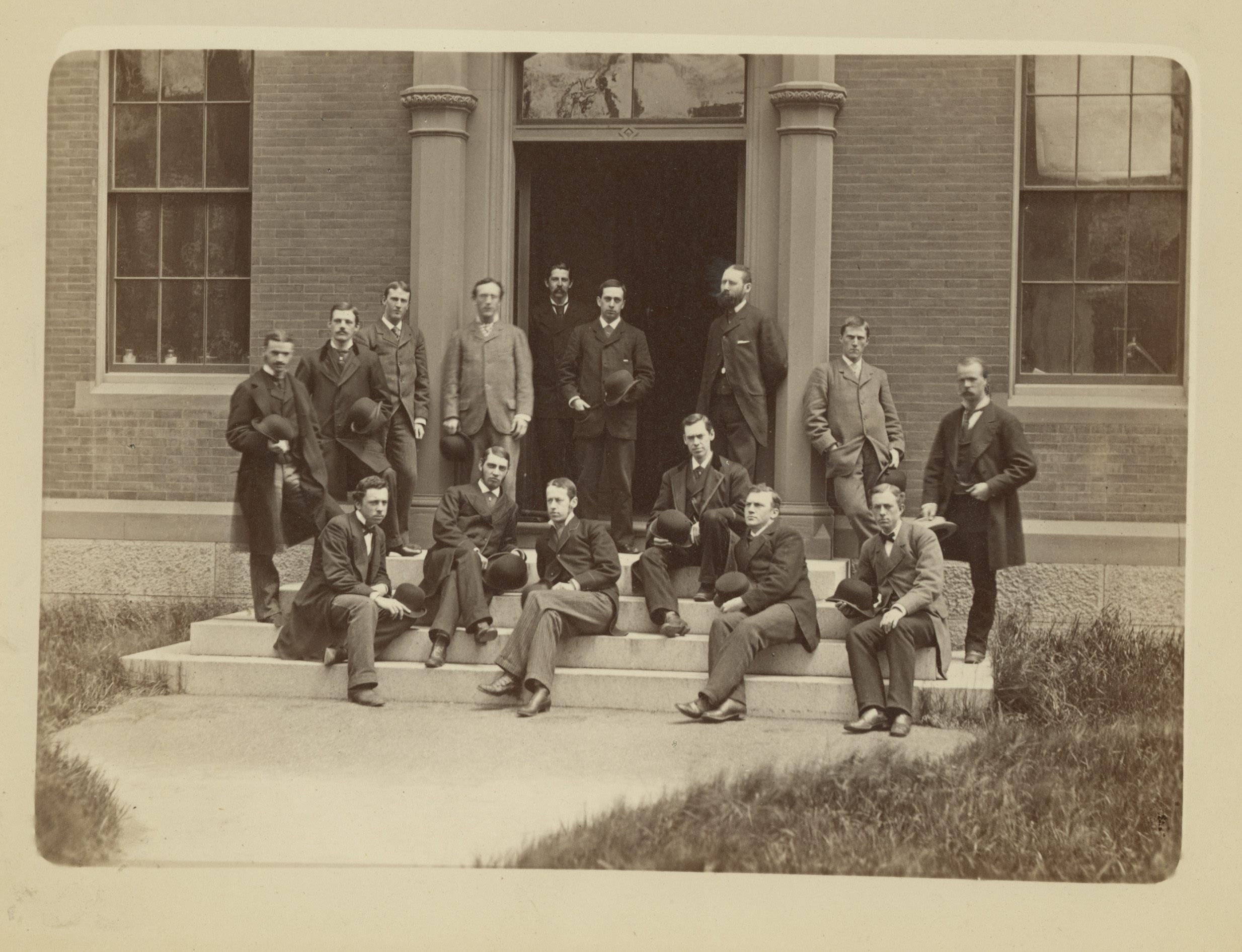
his clasmates at Harvard Medical School circa 1881. Huse is
sitting on the steps of HMS in the center of his class
photograph.

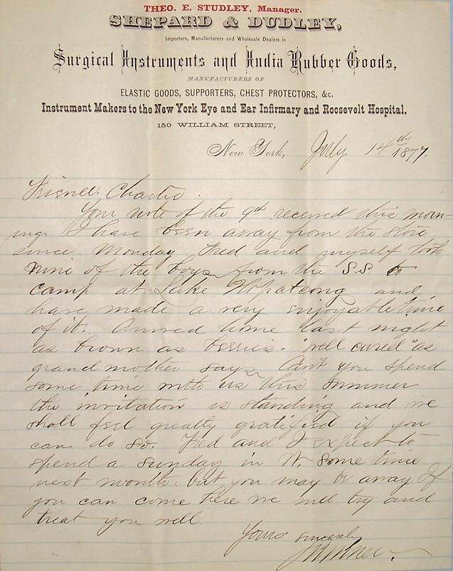

Displayed
are Dr. Huse's Cammann stethoscope marked Shepard & Dudley,
N.Y., and surgical instrument kit, obstetrical forcep and urethral
dilators all marked Shepard & Dudley, New York. Also shown is a
letter from the proprietor of Shepard & Dudley, New York,
dated July 14, 1877, to Friend Charles [Huse] asking if he
would like to join their family for a summer vacation. The letter
implies that Charles knew them because he purchased the
medical equipment that he intended to use as a medical
student from their store.

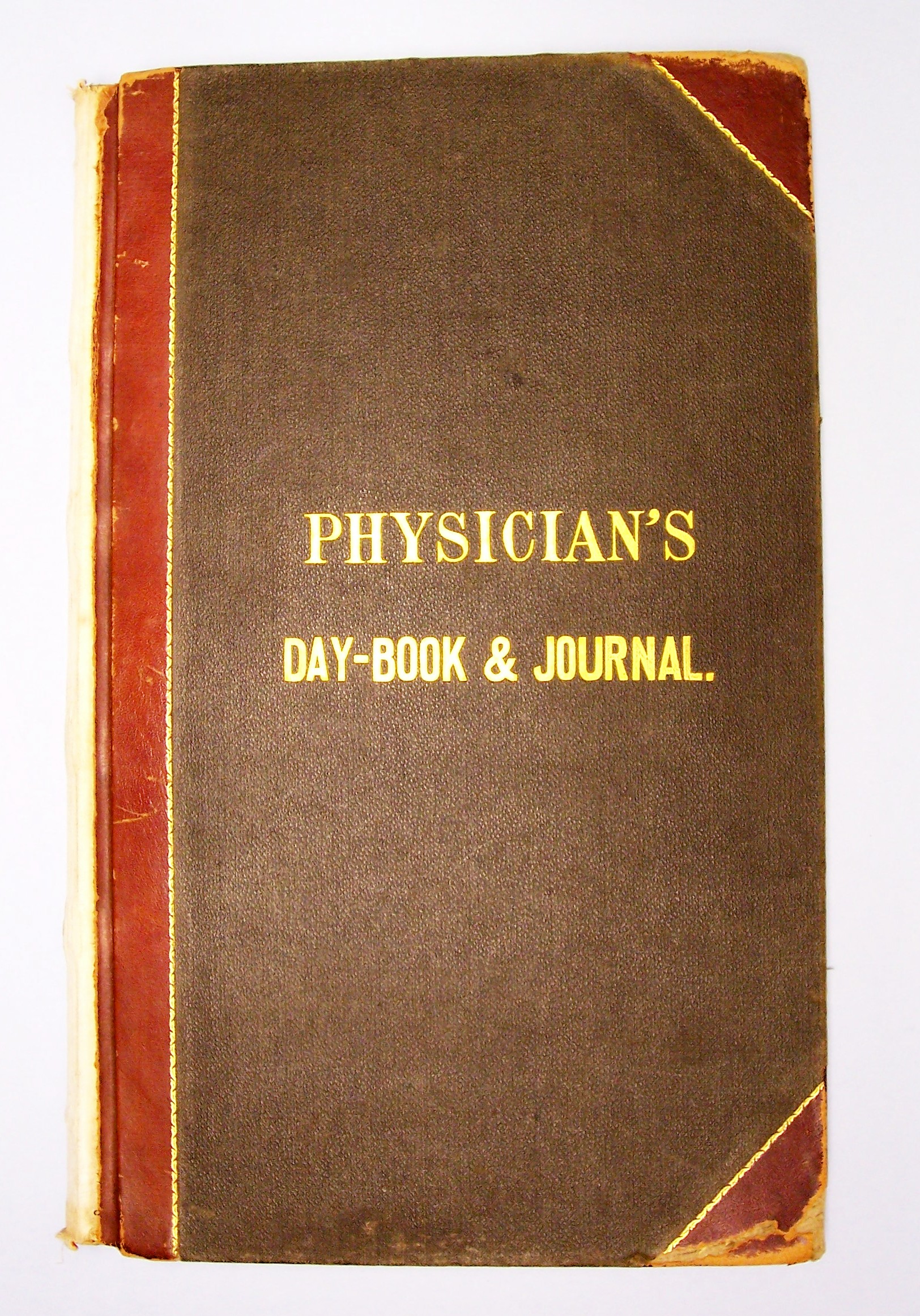
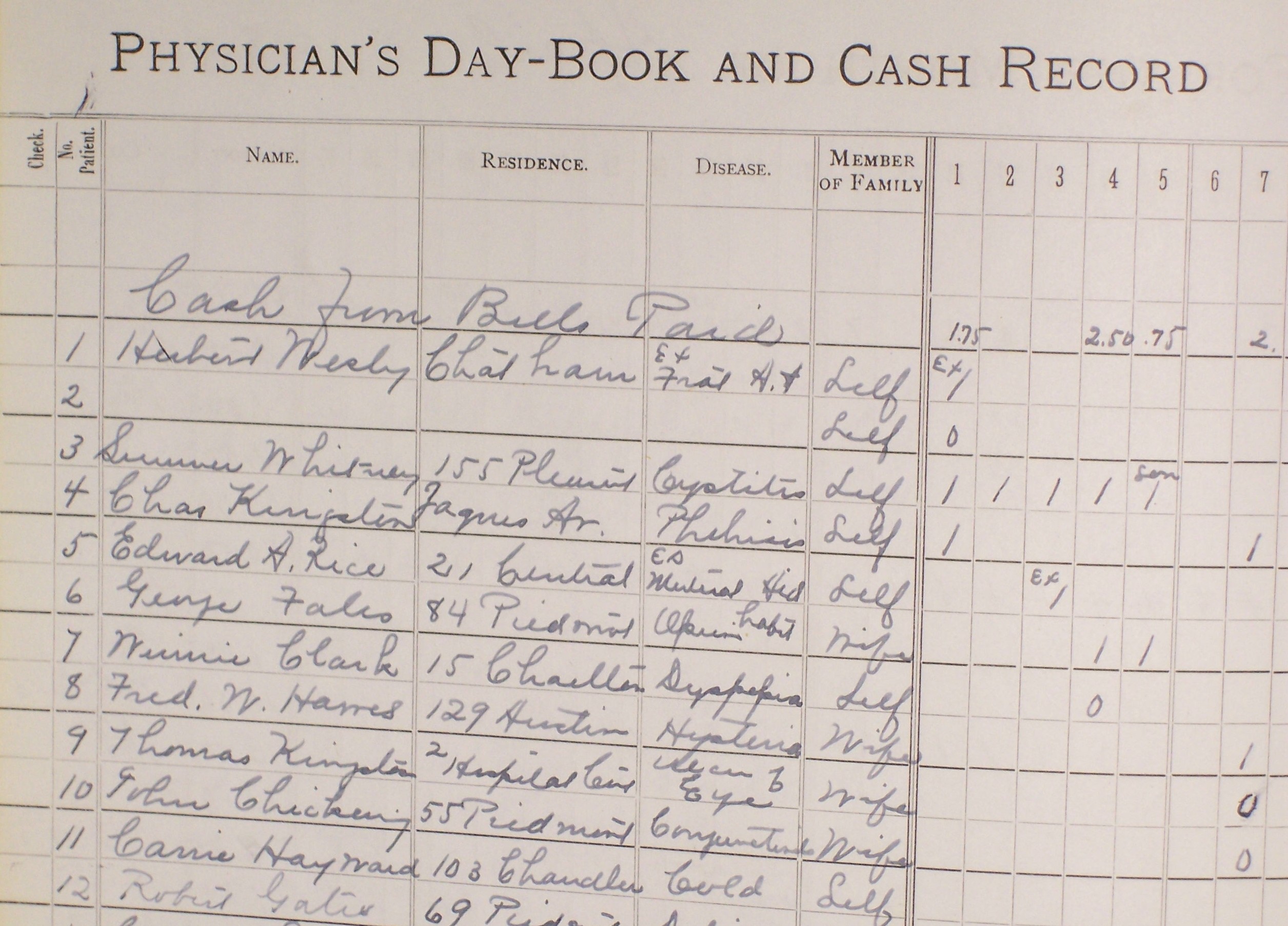

Dr.
Huse kept meticulous records of each of his patients in his
Physician's Day-Book & Journal shown above. Note patient # 7
on page 6 displayed to the left and in a close up of the page
on the right, who Dr. Huse diagnosed as having Dyspepsia on
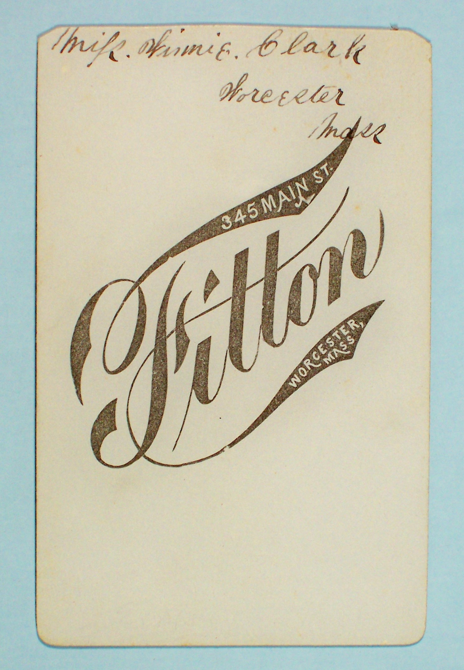
May 4, 1883. This patient was Miss Winnie Clark whose carte-de-visite
albumin photoghraph, circa 1880, is shown with her wearing a
beautiful Victotian mohair wool hat and ornate gold chain. Hand written
in old stlye dip pen ink on the back of the cdv is
Miss.Winnie.Clark / Worcester Mass, as well as the backmark of
the photographer: Fitton, 345 Main St., Worcester, Mass.

Dr.
Huse's records included individual bills for each patient, a copy of
each bill and a Physician's Hand-Book to keep track of payments. A
routine visit to the doctor cost $1.00 !
THE NAMMACK STORY
Bellevue
Hospital in New York City is the nation's oldest public
hospital. It was founded in 1736 as the Alms House, a six bed
infirmary for the poor, and was located at the site of the current
City Hall in lower Manhattan. In 1794, the Belle Vue farm on
Manhattan's East Side was used to quarantine victims of the yellow
fever epidemic in New York City. In 1811, the Kips Bay farm, just
north of Belle Vue, was purchased by the city for a larger almshouse.
McKim, Mead & White designed the Bellevue Hospital buildings
between 1908 and 1939 that stand today on the land of the Belle Vue and
Kips Bay farms on First Avenue and 27th Street. The Bellevue
Medical College was established in 1861 and the first School of Nursing
in the nation based on Florence Nightingale's philosophy was opened in
1873 at Bellevue. In 1841, the University Medical College was
established as the Medical Department of the University of New York on
First Avenue and 26th Street. The College changed its name to New York
University in 1896. In 1898, Bellevue Medical College and
University Medical College of New York University consolidated as the
University and Bellevue Hospital Medical College, ultimately changing
its name to New York University School of Medicine in
1960. Bellevue Hospital remains today the
nation's premier public hospital and continues its long
tradition of serving as a primary teaching hospital of NYU Medical
Center.
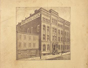


On
the left is a photo of the Unversity College of Medicine (NYU),
1886. On the right is a photo of Bellevue Hospital, 1886.
These
photos are taken from the "University of the City of New York / Medical
Department / Forty-Sixth Annual Announcement of Lectures and Catalogue
/ Session 1886-87 /New York: 1886." In the middle is a view of Bellevue hospital on its 275 birthday in 2011.
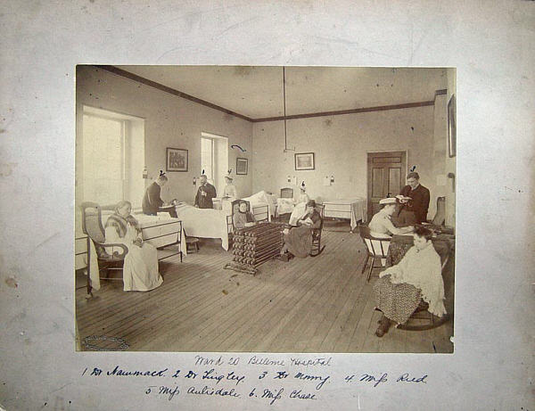

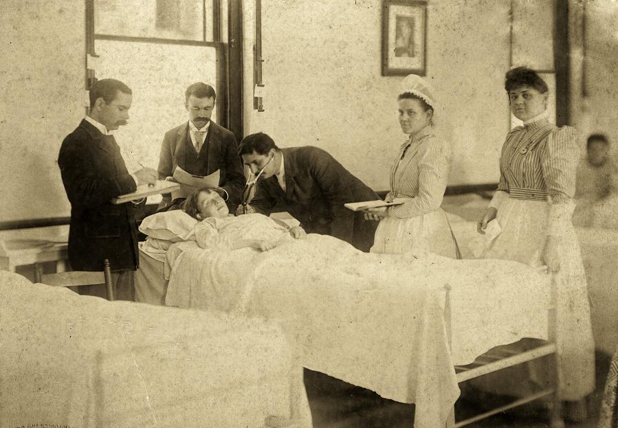
Photo of Women's Ward 20 at Bellevue Hospital in
New York City, circa 1888.
The
photo has a legend identifying the hospital, ward, physicians and
nurses bearing the mark of the Bellevue Hospital Photography
Department, NY City. The enlarged photo on the right shows Dr. Nammack
holding a Cammann stethoscope in his left hand, Dr. Tingley taking
notes, presumaby of Dr. Nammack's ausculatory findings and nurse Ried
with an open chart. Note that Dr. Nammack is also holding his patient's
hand, perhaps to reassure her, as he discusses his observations.
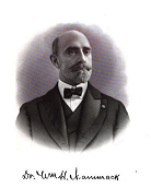
Photo of Dr. Wm. H. Nammack, circa 1900, from the book A
History of Long Island by William S. Pelletreau.
Dr.
William H. Nammack was a 1886 graduate of Bellevue Medical College and
was one of four "successful candidates for appointment in the
Bellevue Hospital ". He served as a House Physician on the 3rd Medical
Division at Bellevue Hospital in 1886-1887. In September of 1887, Dr.
Nammack was appointed the Medical Superintendent of the Insane
Pavillion of Bellevue Hopsital. He remained a member of the outside
hospital medical staff for another nine years. Dr. Nammack was a
physician and surgeon who originally practiced general medicine at 11
Rutgers Street in New York City. In 1892, he established his home
and surgical practice in Far Rockaway. Dr. Nammack was visiting
physician and pathologist at St. Johns's Hospital in Long Island City,
and aslo served as the Coroner of Queens for a number of
years. He was a memeber of the Bellevue Hospital Alumni Society, New
York State and County, Queens County and Nassau County Medical
Associations, and Society for the Relief of the Widows and
Orphans of Medical Men. His son Griswold D. Nammack, grandson
Griswold P.D. Nammack, brother Charles E. Nammack, and nephew Charles
H. Nammack were also all physicians affiliated with Bellevue Hospital.
Dr. Nammack's great grandson Thomas W. Nammack is the Headmaster of The
Montclair Kimberley Academy, Monclair, New Jersey, and provided part of
this history of the Nammack family.
Dr. Witter Kinney Tingley
was also a graduate of Bellevue in 1886 and another of the four
graduates to serve as a House Physician on the 3d Medical
Division in 1887-1888. Dr. Tingley practiced General Medicine in
Norwich, Connecticut and served as President of the Norwich Medical
Association in 1890. He was one of the incorporaters of and Visiting
Physician to Backus Hospital in 1892.
Dr. Robert Alexander
Murray was a graduate of University Medical College (NYU) in 1873 and a
House Physician on the 2d Medical Division from 1874-1875. Dr. Murray
practiced General Medicine and specialized in Disease of Women at 235
West 23d Street , New York City. He served as Attending Physician,
Diseases of Women, at the Northwestern Dispensary, New York City,
from 1876-1883 and Assistant Professor of Obstetrics at University
Medical College (NYU) from 1876-1886. He also served as an Instructor
in Obstetrics, New York Polyclinic from 1887-1888. Dr.
Murray authored various articles on obsterical subjects in medical
journals.
Ms. Ried, Autisdale and Chase were Nurses at Bellevue Hospital.
Another
photo of Rounds at Bellevue Hospital from the Bellevue Hospital
Photography Department, circa 1891. Similar to the previous
photo, a physician is using a Cammann Stethoscope to auscultate
the heart of a young patient, while a medical student takes
notes of the physical diagnostic findings and a nurse holds an open
chart.
( Photo courtesy of the NYU School of
Medicine Archives )
THE WHITE COLLECTION
Eclectic medicine was a branch of American medicine which made
use of botanical remedies along with other substances and physical
therapy practices, popular in the latter half of the 19th and first
half of the 20th centuries. "Eclectics" were doctors who practiced with
a philosophy of "alignment with nature," learning from and using
concepts from other schools of medical thought. They opposed the
techniques of bleeding, chemical purging and the use of
mecury compounds common among the "conventional" doctors of that
time.
The Eclectic Medical Institute in Worthington, Ohio graduated its first class in 1833. After the notorious Resurrection Riot in 1839, the school was evicted from Worthington and it settled in Cincinnati during the winter of 1842-3. The Cincinnati school, incorporated as the Eclectic Medical Institute (EMI), continued until the last class graduation in 1939 more than a century later. Over the decades, other Ohio medical schools had been merged into that institution. The American School of Medicine (Eclectic) in Cincinnati operated from 1839 to 1857, when it merged with the Eclectic Medical Institute.
Eclectic Medicine expanded during the 1840s as part of an large, populist anti-regular medical movement in North America. It used many principles of herbal medication but chose to train doctors in physiology and more conventional principles, along with botanical medicine. The American School of Medicine (Eclectic) trained physicians in a dozen or so privately funded medical schools, principally located in the midwestern United States. By the 1850s, several "regular" American doctors, especially from the New York Academy of Medicine, had begun using herbal salves and other preparations.
The movement peaked in the 1880s and 1890s.
The schools were not approved by the Flexner Report (1910), which was
used to decide on accreditation of medical schools. By World War I,
states were adopting curriculum requirements that followed those
articulated by the AMA. Those schools preferred pharmaceutical
medicines to botanical extracts.This effectively forced the Eclectic
Medical Schools to either adopt the new model or fold. Although the
last Eclectic Medical School closed in Cincinnati in 1939, modern
medicine now recognizes "alternative" medicine as a potentially
important adjunct to "traditional" medical therapy
Dr. Harvey A. White was a 1882 graduate of Eclectic
Medical Institute (College) in Cincinnati. After
graduation, Dr. White practiced in
Missouri in 1882, Illinois in 1883, including in Chicago where this
photo was taken in 1904, and White Plains, New York in 1909. He retired
to Tampa, Florida where he died in 1917. Dr. White's Cammann
binaural stethoscope is shown to the right of his photo.
Prior to becoming a doctor, Harvey A. White was a soldier in the
civil war. He enrolled on December 17, 1861 as an artificer in the
newly formed Battery E of the First Illinois Regiment of Light
Artillery commaned by Captain A.C. Waterhouse in the 5th Division under
General William T. Sherman as part of the Army of the
Tennessee commaned by General Ulysses S. Grant. Battery E played a key
part in one of the boodiest battles of the civil war at
Shiloh, Tennessee. In March of 1861, Grant moved his
Union Army up the Tennessee river into west Tennessee around
Shiloh in preparation to attack and seize the Corinth rail junction
that connected the eastern and western Confederate states, hoping to
deal a fatal blow to the Confederacy and end the war quickly. General
Albert S. Johnston also realized the strategic importance of the
Corinth rail junction and poistioned his newly christened Army of the
Mississippi to launch a suprise attack on Grant's forces. As an
artificer, Harvey White could have been in the forward
position with two artillery guns of Battery E confronting the
initial suprise attack by Johnston's Confederate Army on
April 6, 1862. The Confederate forces overran the Union
Army's frontlines and pushed back Grant's forces back to their last
line of defense at the sunken road, later described by a Union soldier
as a "hornets nest" because of the sound made by the constant rain of
bullets fired in that battle. The casualties on the first day of battle
were immense,
including the dealth of the confederate leader, General Johnston.
General Grant was determined to counterattack rather than retreat. On
April 7th with the arrival of General D.C. Buell's Army of the Ohio,
Grant launched an aggressive counterattack defeating
the Confederate forces on the second day of battle. The two
day battle was costly. More Americans fell at Shiloh than the
total casualties in all of the previous wars
fought by the United States. According to the official records 23,746
men were killed, wounded or missing and both sides now realized that
this would be a long and bloody war, just as Dr. Robinson had predicted
in his letter to his father written the previous year on April 28,
1861 (see Dr. Robinson's story in the previous page).


Shown above in the top figure is the 1890 Prang and
Thulstrupt print of the Battle of Shiloh of Union forces last line of
defense at Hornet's Nest at 9am on April 6, 1862. The photo on the left
is the forward position of the two guns of
Battery E, First Illinois Light Artillery with a marker of the
site of the initial battle of Shiloh at 6am on April 6th. The
plaque on the marker
reads "Waterhouses's Battery, 'E' commaded by 1. Captain Waterhouse,
Wounded. 2. Lieut. A.E. Abbott, Wounded. 3. Lieut. John A. Fitch. Two
guns of this battery were advanced about 300 yards but soon fell back
to this position where the whole battery went into action. This ground
was held from 7:00 A.M. to 9:30 A.M., when the battery lost two
guns and moved back about 100 yards. Its loss in the battle was 1 man
killed; 3 officers wounded and 14 men wounded; total, 18." Harvey White
was given a medical discharge by order of General U.S. Grant at
Monterey, Tennessee on June 10, 1862 for a neck injury suffered in the
battle of Shiloh. The carnage that Harvey White witnessed at
Shiloh may have ultimately lead him to become a doctor.
Several physicians came up with their own ideas for
stethoscopes using materials that were commonly found in a physician's
office. For example, in 1884, E. T. Aydon Smith described a stethoscope
he invented: "..the chest-piece of which is formed by a pair of
ear-specula, the tubes are Jaques' India-rubbercatheters, and the
ear-pieces those of an otoscope."He designed his instrument to be used
as a simple binaural, a differential stethoscope (by using one of the
ear-pieces and chest-pieces for each ear), a tourniquet (by wrapping
the rubber around the arm) and, also as a urinary catheter for both
males and females. Quite a diverse instrument!
Another modification was Dr. D.M. Cammann's modified instrument
(Dr.
George Cammann's son), which incorporated a chest piece with a rubber
ball, which was used as a suction cup to apply it to the chest. This
would leave the hands free to percuss to chest. It was found to be of
little value to the untrained ear.
|
READ DR. CAMMANN'S SON'S ADDRESS REGARDING HIS MODIFICATION OF HIS FATHER'S STETHOSCOPE |
New
improvements to the stethoscope focused on the tension mechanism to
hold the ear-pieces to the head of the physician. There was a design of
a spring-tension mechanism which had a screw that could be turned to
open or close the ear-pieces. The first of these new models was
KNIGHT'S model, which had a characteristic design on the poles that
attached to the ear-tubes. The Charles Truax catalog of 1890 states that
"Knight's stethoscope does not differ materially from the pattern of
Cammann. The principal change is in the form of the spring, which in
this case is spiral, acting on two levers in the form of a toggle
joint."
The screw mechanism became a great success
and would be included in the models that followed for many years.


On the left
is a late 1860s Cammann's binaural stethoscope with the
additon of an adjustable tension mechanism to the ear tubes that was
described Dr. Frederick
Irving Knight of Harvard. The instrument is known as
Knight's binaural stethoscope. However, the credit for
this design belongs to Moses G. Farmer as Knight referenced
in his article in the Boston Medical Surgical Journal in 1869. The
metal work is German silver, the flexible tubes covering is woven silk,
the bell and junction are ebony, all characteristics of an early
Knight's double stethoscope by the original maker Codman &
Shurtleff, Boston. To the right is a Knight's
Stethoscope with two bells and a Flint Pleximeter and Percussion Hammer
that is shown with its original wood carrying box, Codman &
Shurtleff, Boston, circa 1880. On the left the instruments are shown
out of the box and on the right placed inside the box. The bells were
interchangeable in that each could be screwed on and off the body of
the stethoscope depending on the choice of the physician.
The larger bell was used for auscultation of the lungs and smaller bell
used to localize heart sounds. Note the metal work is now plated
silver. (Photo on left
courtsey of Alex Peck)


Bartlett's Stethoscope shown on the
left was a heavier version of the Knight model, circa 1880.
Bell's Stethoscope, circa 1875, has an all metal chest piece with
rubber rim in this adaptation of the Knight model.
The Cammann
stethoscope on the right has an unusual horn bell, brass ear
tubes and ivory ear tips and is marked H.G. Kern, Philadelphia,
circa 1860.
There
were many different modifications to Cammann's original instrument.
Scott Alison came up with a 'Differential Stethoscope' that consisted
of two independent chest pieces, and was designed to allow the listener
to compare the sounds of two areas of the chest. This instrument was
also found to be impractical because it muffled certain sounds. Most
references list this piece as circa 1885, but it was actually first
illustrated in the John Weiss & Son catalog in 1863.

Alison's
Differential Stethoscope is displayed on the left, circa 1850. The
close up shows the ebony bell chest pieces. It is signed "Ferguson."
Mr. David Fergusson of Giltspur Street, London was the well known
instrument maker for the Bartholomew's Hospital in London and
introduced many of the new instruments that were used in the hospital.
On the far right is an example of a modern
differential stethoscope with metal diaphragm chest pieces,
circa 2000 and next to it is a differential Stethoscope with
plastic bell chest pieces, circa 1930.
In
1885, Charles Denison came out with an entirely new model, based on
Bartlett's. His was based on the idea of funneling sound to the ears,
much like the monaurals. His earpieces were made of hard rubber which
led into woven tubes and a large chest piece. It came with three
interchangeable chest pieces for hearing different types of sounds. The
tension between the earpieces was accomplished by a clever screw
mechanism.Denison's model was widely accepted. So widely,
that many makers began marketing inferior pieces of the "Denison
Stethoscope." Denison became outraged at the poor quality of
stethoscope being manufactured with his name. He delivered a powerful
speech in which he stated precisely how his stethoscope was to be made,
condemned the makers of poor instruments, and praised one American
company for their quality craftsmanship.

Denison's Stethoscope, circa 1885.
Left example with multiple size bells, courtesy
of the Mutter Museum. Right
examples are in this collection, shown without and with large bell. The
stethoscope was used withhout the bell for auscultation of
children.

Davis modification of the Cammann Stethoscope which incorporated a wire metal tension spring between the metal ear tubes. In this early model, the wood chest bell was attached to the ear piece by a flexible tube which inserted into the wooden ball connecting the ear piece, circa 1880. On the right is a later model Davis stethoscope with simple bell and metal tension mechanism between ear pieces, circa 1890 .

On the left is a Matthews stethoscope with
detachable chest piece and ear tubes for easy portability, circa 1882.
Shown on the right is a very
unusal folding stethoscope with ivory ear pieces and a unique bell that
is a smaller version of the original Cammann bell with a insertable
plug designed after the William's plug, circa 1890. This two piece bell
is made of wood and allowed the auscultator to change the size of the
bell by inserting or removing the plug.
Convenience
came in several ways. Longer tubes added more flexibility, but other
methods were used as well. Around 1880, Lynch's stethoscope was
marketed, which folded onto itself and thus greatly reduced it's
length. Sheppard's model (1890), by contrast, folded the earpieces
together reducing the width of the instrument.


Lynch's folding stethoscope, circa 1880. This piece folds top to bottom as shown on the right. Sheppard's folding stethoscope in its original box shown on the right. This piece folds in half. On the left is the top of the cardboard box which is marked "Sheppard's Stethoscope / patented July 7th, 1890."
The
next landmark improvement was the invention of the 'Ford's Bell' chest
piece in 1885. This simple model was made of steel with either
gutta-percha, ebony, or even ivory at the base. It funneled sound into
two tubes, usually made of rubber, which led to the earpieces.

Three
examples of early Ford's bell stethoscopes with a simple metal band
tension mechanism holding the ear tubes together. On the left
the Ford's bell is made of ebony, circa 1890. In the middle the Ford's
bell chest piece is made of ivory with,circa 1895. On the right the
Ford's bell is made of metal, circa 1895.
Three examples of binaural stethoscopes
with interesting tension mechanisms to hold ear tubes together.
On
the left is a Down Brothers stethoscope with finger rest, interesting
screw spring tension mechanism, ivory ear tips and Ford bell made of
ivory, circa 1900. The ivory bell unscrews to creat a smaller bell for
the examination of children. In the middle is a binaural stethoscope
marked Gallante, circa 1890. The wood chest piece and ear tips are
typical of French binaurals of this period. Note the spring tension
mecahnism between the ear pieces. The right photo shows a Holborn
stethoscope with a slender, wood bell typically used for
children, Herschel's locking mechanism with lever and ivory ear
tips, circa 1900.


The left three examples areDown Brothers
stethoscopes with Ford's bells made from differnet materials.
On
the left the stethoscope has a finger rest and Ford bell made of
aluminum, circa 1920. In the middle is a folding stethoscope with the
mark of the "British Army" with ivory Ford bell and wood ear
pieces, circa 1910. On the right is a folding stethoscope with an
anti-chill, rubber cushioned Ford bell, circa 1910.


Binaural stethoscopes with flexible rubber
tubes to connect the ear pieces to the chest piece, circa 1880.
The
example on the left is Chinese and has ivory ear pieces and a
reversible ivory chest piece. Each end of the reversible chest piece
was a different size and shape, presumably to examine different parts
of the body. The middle ivory example is Japanese. The one on the right
has a gutta percha chest piece.

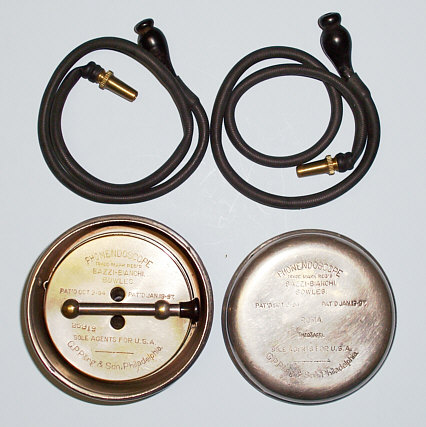
Examples of two American Phonendoscopes made by G.Pilling & Co. On the left the phonendoscope is in its original velvet lined, wood box, circa 1909. The photo in the middle is another phonendoscope in its original metal case with the chest piece in the case and its extension screwed onto the top for carrying. To the right the stethoscope is put together for auscultation with the ear pieces made of gutta-percha attached by flexible tubes with metal ends to the chest piece and the extension tube screwed into the diaphragm of the chest piece.


Three examples of phonendoscopes made in Europe. On the left is a model with an adjustable valve to vary the intensity of sound made by Osker Skaller A.G. of Berlin. In the middle is a German model with gutta percha ear pieces and on the right a similar French model with glass ear pieces, circa 1900. All the phonendoscopes are displayed in their original, velvet lined, leather covered,carrying cases.


Schreiber's Stetheskop made of metal,
circa 1900.
As
shown from left to right, it could be set up as a monaural or by
sliding the chestpiece down the stem to expose the shaft, a
phonendoscope chestpiece could be attached to the stem. A close up of
the phonendoscope chestpiece is shown on the far right.


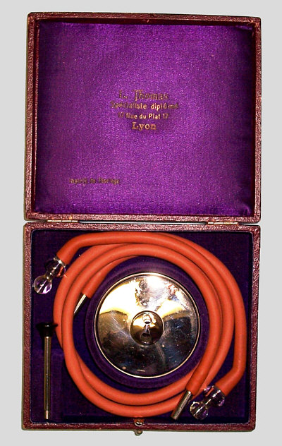
Wincarnis
stethoscope, circa 1890. On the right the stethoscope chest piece is
shown in its original case. On the left the flexible tubes with ivory
ear pieces and the chest piece taken apart are displayed. Note the
plastic diaphragm against which either of the two wooden disks would
rest
Marsh's Stethoscope, or
Marsh's Stethophone, is a very interesting invention. It comes with a
small dial on the back which has a pointer and the letters 'L,' 'S,'
and 'W' engraved on it. These stood for 'Loud,' 'Soft,' and 'Weak,'
respectively. The examiner would dial in the type of sound he was
listening to in order to be able to hear it better.

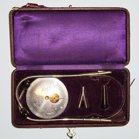


Marsh's Stethophone, circa 1896
This is one of two examples known. The other is in the collection of
the Armed Forces Institute of Pathology.

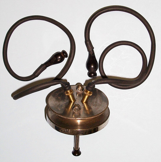
Magniphone Stethoscope, circa 1900.
The piece below is very interesting in that it combines the binaural ear piece of a stethoscope with a very long tube and wide bell charcteristic of a hearing aid or conversation tube. In fact, this piece is neither a stethoscope or hearing aid, but rather a "re-educative binaural tube to enable the patient to read or speak to himself and thus stimulate the dormant auditory centre by natural means of the voice" in order to treat middle ear deafness (Mayer&Phelps, circa 1931).
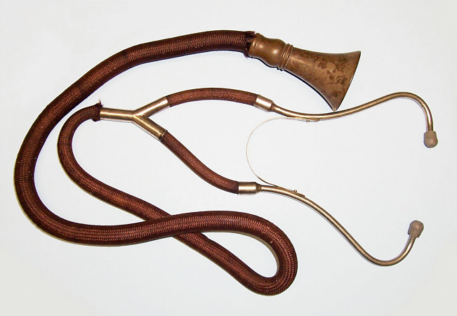
Re-educative scope, circa 1931.
We are always interested in acquiring new items for the collection. If you have any items for sale or question please do not hesitate to contact us.