
Click on IMAGE to enlarge photos. Click on BLUE link to read articles.
Assessing the sounds of the human body was reported in the ancient
medical literature. Amongst the earliest known medical manuscripts are
the medical papyruses of ancient Egypt dating to the seventeenth
century B.C., which referred to audible signs of disease within the
body. Hippocrates, the Father of Medicine, advocated for the search of
philosophical and practical instruments to improve medicine in 350 B.C.
He discussed a procedure for shaking a patient by the shoulders
(succussion) and listening for sounds evoked by the chest. Hippocrates
also used the method of applying the ear directly to the chest and
found it useful in order to detect the accumulation of fluid
within the chest. In the sixteenth century, the renown surgeon Ambroise
Pare noted that "if there is matter or other humors in the thorax, one
can hear a noise like that of a half filled gurgling bottle." The
distinguished scientist William Harvey, in his 1616 lecture on the
structure and function of the heart, described the heart's motion as
"two clacks of a water-bellows to rayse water" and noted that "with
each movement of the heart, when there is delivery of a quantity of
blood from the veins or arteries, a pulse takes place and can be heard
within the chest." The French physician Jean-Nicolas Corvisart, who is
considered the founder of French clinical medicine, was accustomed
to placing his ear over the cardiac region of the chest to listen to
the heart. Bayle and Double, who like Laennec were students of
Corvisart, used the unaided ear to listen to the heart of their
patients. Double suggested the regular use of this technique in his
treatise on Semiologie published in early 1817, prior to the
publication of Laennec's
treatise on auscultation. He wrote "the ear should be brought against
the thoracic wall" in order to appreciate the noises inside.
Nevertheless, the evolution from listening
with
the unaided ear (immediate auscultation) to the aided ear (mediate
auscultation) awaited Laennec's invention of the stethoscope.

An engraving of a physician examining a patient by
"immediate" ausculatation, in which the doctor placed his ear on the
chest of the patient to hear the sounds made by the lungs during
breathing.The print shows a group of phiscians, medical students and
nurses observing the physician performing his exam. The print
is entitled "A Visit to the Hospital" by the artist Luis Jimenez
Aranda. It was copyrighted in 1894, and originally displayed at the
Chicago World's Fair in 1893.
RENE LAENNEC: INVENTOR OF THE STETHOSCOPE
The
stethoscope was conceived in 1816 when a young French physician named
Rene Theophile Hyacinthe Laennec was examining a young female patient.
Laennec was embarrassed to place his ear to her chest (Immediate
Auscultation), which was the method of auscultation used by
physicians at that time. He remembered a trick he learned as a child
that sound travels through solids and thus he rolled up 24 sheets of
paper, placed one end to his ear and the other end to the woman's
chest. He was delighted to discover that the sounds were not only
conveyed through the paper cone, but they were also loud and
clear. The onset of Laennec's stethoscope research began in 1817 at the
Necker hospital. The first published oberservation documenting
auscultation
using the stethoscope (Mediate Auscultation) was in March 8,
1817, when Laennec noted examining a 40 year old chambermaid,
Marie-Melanie Basset. Laennec's research activities about mediate
ausculatation were first brought to public attention with his
consultation in June of 1817 for Mme de Stael, who was the daughter of
the Neckers and an author who criticized the rise of Napoleon's empire.
Her personal physician described Laennec's consultation: "Another well
known doctor [Laennec], using a horn of paper, which he placed with one
end on a part of the thorax and the other in his ear, believed he
diagnosed a hydothorax, an could even hear a sort of undulation. One
can well understand that I considered this method of investigating the
inte rior of the chest to be very strange, and I did not share his
opinion, in spite of the regard I might have for him."
rior of the chest to be very strange, and I did not share his
opinion, in spite of the regard I might have for him."
Laennec began his study of medicine under his uncle who was
a
professor of the faculty of medicine in Nantes. Eventully he entered
the Paris University where he studied medicine under Jean-Nicolas
Corvisat, who was a proponent of immediate ausculatation and
percussion. He was a devout Catholic and his charity to the poor was
proverbial, taking him to the Hospital Necker after his studies to care
for the sick and poor. It was at the Hopital Necker that Laennec
invented the stethosopce. His clinical work allowed him to correlate
auscultation and post-mortem findings thus defining
disgnoses of diseases of the chest. On February 23, 1818, Dr. Laennec
presented his findings in his Memoire sur L'Ausculatation to the Academy of Medicine in Paris, later
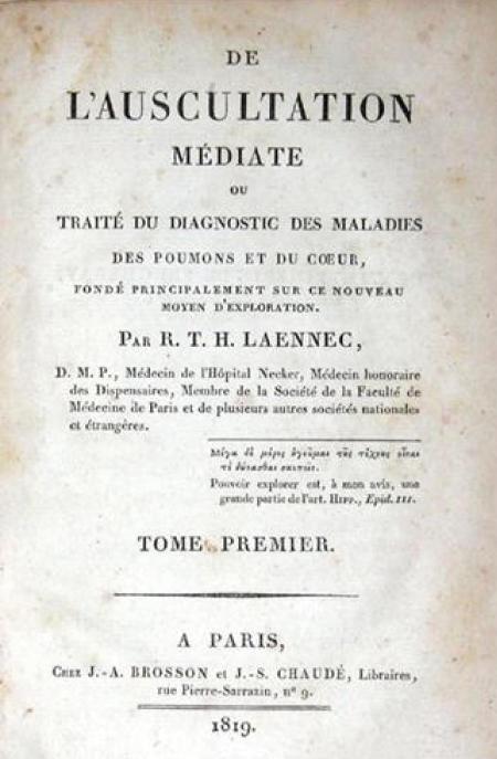
publishing his comprehensive treatise De L'Auscuatation Mediate of his work in 1819. In 1823,
Laennec was appointed professor of clinical medicine at Paris
University resigning from Necker hospital to preside over his former
mentor Corvisat's clinique interne at the Charite hospital. A few years
later just prior to his death he bequeathed his own stehoscope to his
nephew, Dr. Meriadac Laennec, referring to it as the "greatest legacy
of my
life." It is said that Meriadac diagnosed Laennec as having
tuberculosis using his stethosocpe. In his lecture on Laennec in 1883,
the preeminent American authority on ausculataion, Dr. Austin Flint
said "Laennec's life affords a striking incidence
among others disproving the vulgar erorr that the pursuit of science is
unfavorable to religious faith."
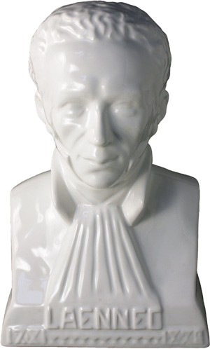
The
faience buste shown in the middle photo was created by Georges Robin,
HB-Henriot Quimper, in 1926 for the centennial of the death of Docteur
Laennec. This reissue white enamel monochrome from the original mold
has a height of 12.6 inches and is number 15 of a limited series of 100
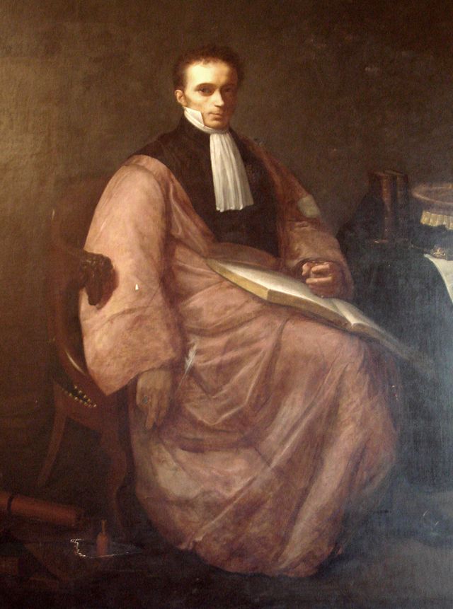
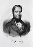
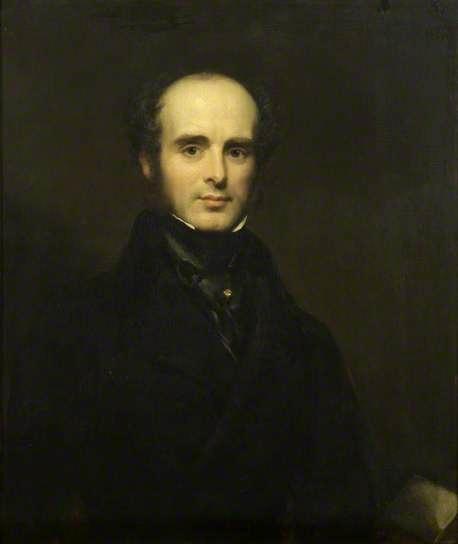
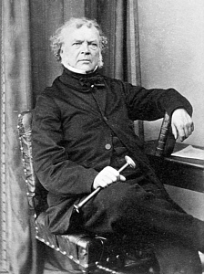
pieces. The buste was based on the the oil portrait
on the left of Rene Theophile Hyacinthe Laennec by
Paul Dubois,
circa 1854. This posthumous
portrait was comissioned by the Medical Faculty of the Universite Paris
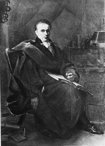
Descartes and painted from the only full length portrait of Dr. Laennec shown in the
photographic print on the right by Alexandre Dubois, a struggling artist
who painted Laennec as payment
for his medical services in 1812. The original portrait did not dispaly
Laennec's stethoscope and the family had the artist add the stethsocpoe to his portrait in 1825 after the invention of
the instrument by Dr. Laennec in 1816.
Note
Laennec's stethoscope shown in the lower left hand corner of both
portraits. The 1854 Paul Dubois painting changed Laennec's garb from
the 1812 Alexandre Dubois original portrait, perhaps because the
Faculty of Mediciane wanted to show Laennec in a their traditional faculty
robe. In the 1854 portrait, Laennec is now wearing an academic robe
with a matching hat on the table instead of a lamp, as well as two
matching books on the table instead of the large book
dispalyed in the original version. Laennec's treatise on asculatation
was publised in two volumes.
( Photo on the left courtesy of Museum of the History of Medicine, Paris University Descartes and on the right from the Wellcome Museum, London)
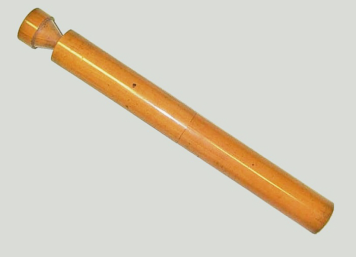


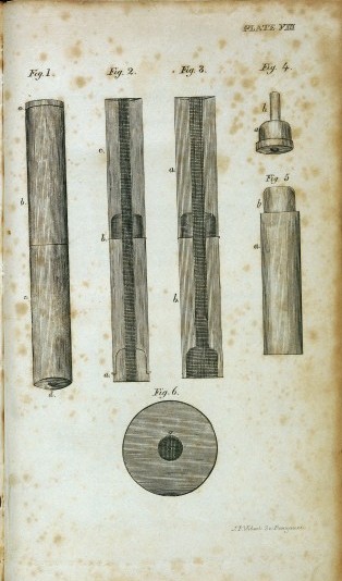
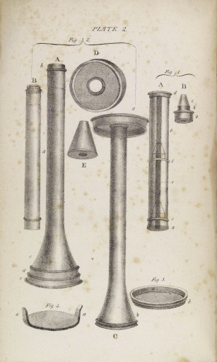
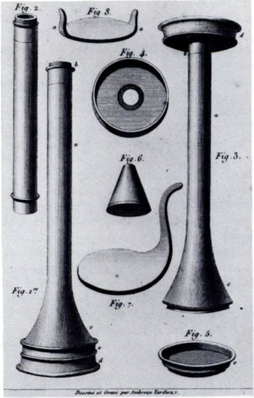
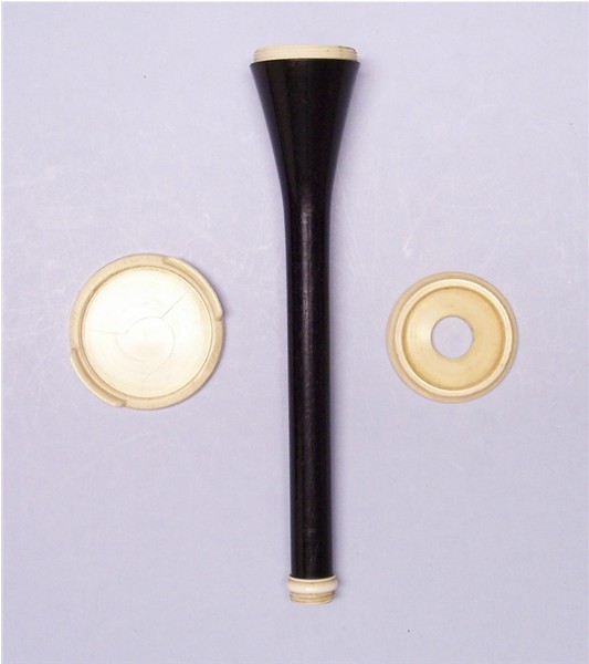
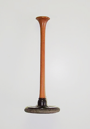
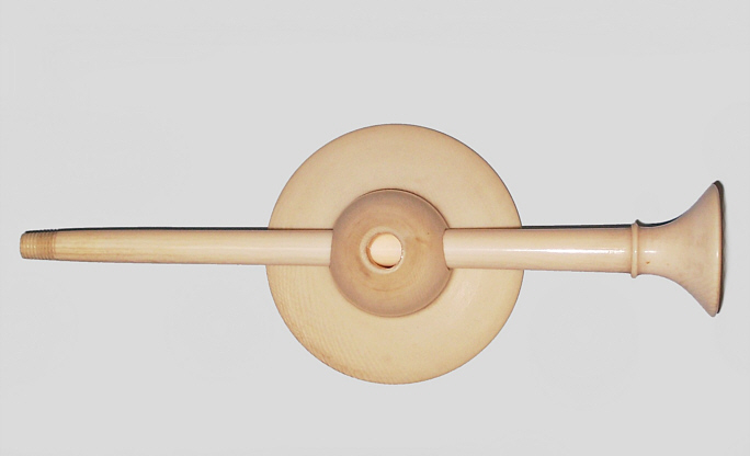
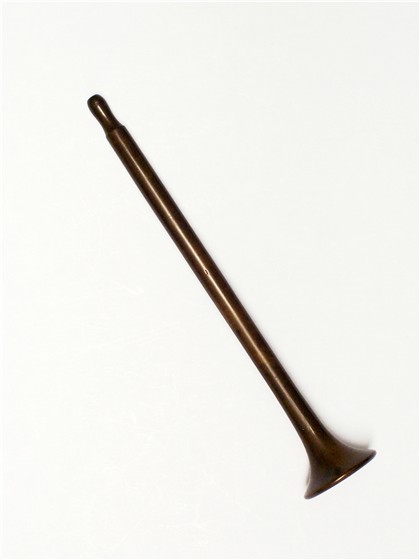
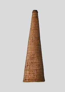
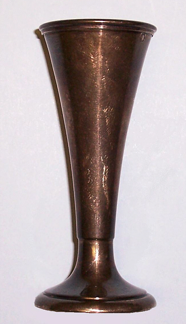
Original version of the
Laennec stethoscope c. 1817 made from boxwood and turned by Laennec. It is not suprising that Laennec used boxwood as one of the early woods he tested to make stethoscopes, because as a
flutist he knew that flutes were usually made of European
boxwood, a finely grained, light
colored wood that is very good turning material and carries musical
sounds very well. This cylindical stethoscope is made with
three parts fitting together by wood screw thread and brass tube
fitting with an overall length of 12.6 inches and a diameter of
1.5 inches. Both ends are slightly concave. This first version is
illustrated in Laennec's first edition 1819 text on auscultation
which
described the stethoscope as having an overall length of
12 inches and a diameter of 1.5 inches. Laennec turned the first
stethoscopes himself and these were somewhat longer than described in
his text. The stethoscope in this collection shown above has the same
features as a
surviving stethoscope that Laennec also made and gave to his friend
Professor Lobstein of Strasbourg (www.woodlibrarymuseum.org)
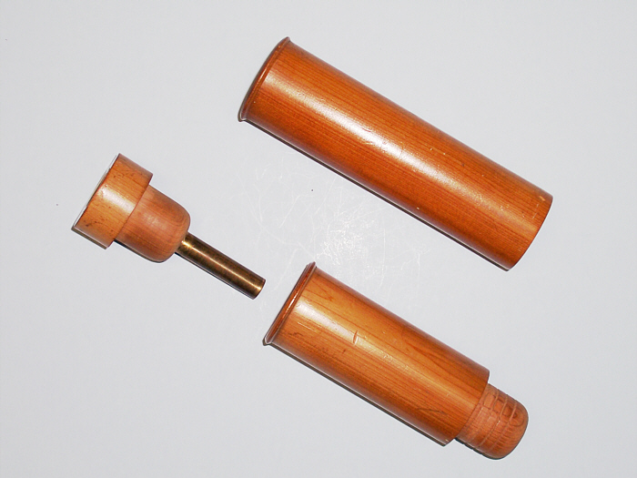
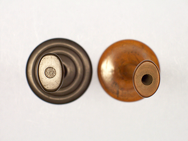
On the left
the stethoscope is assembled with the chest plug protruding
from the funnel shaped chest end of the stethoscope. On the right
the stethoscope is taken apart revealing the wood screw thread that
attaches the two parts of the body of the stethoscope and the chest
plug with brass tube fitting that holds the chest plug in place in the
funnel shaped chest end. Also shown is the title page from Laennec's 1819 text on
mediate ausculatation with the plate illustrating his
stethoscope.
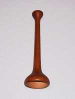
An unusal first version Laennec stethoscope was also made as a short stehtoscope measuring about 8.5 inches long.
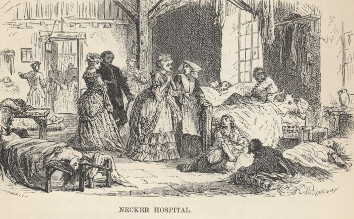
The Necker Hospital
was established in 1778 by Madame Necker, wife of Jacques Necker,
Minister of Finance in the court of Louis XVI. Madame Necker was
appalled at the conditions of Paris hospitals caring for the poor and
opened a 100 bed hospital under her direction and at her own
expense, that still bears her name today in order to
provide a facility that would serve as a model of
efficiency and hygiene for hospitals of Paris. In 1816, Laennec was
appointed physician at the Necker hospital in Paris at which his
studies on auscultation would result in his most important contribution
to medicine.


Shown on the left is a photo of
the Hopital Necker, Rue de Sevres, Paris, circa
1900. Note the large cental court
gardens that were used to grow herbs as sources of medications. In
the center is a photo of the memorial plaque on the outside wall of the
Hopital Necker commemorating Dr. Laennec's discovery of the
stethoscope. The palque reveals a sculptured "cylindre"
of the original model of the stethoscope made by Laennec as
illustrated in his 1819 treatise and a later 19th century
model of a typical monaural stethosocpe. There is also a snake
(typical of a medical caduseus) wrapped around both stethoscopes
and a motar (bowl used to crush ingredients in order to
prepare medications). The plaque is still in place today. To the right of the plaque is a 2011 photo
of the renovated original Necker Hospital with its current courtyard in the foreground. On the far right is a
illustration from "A
Popular history of France" by M. Guizot of a ward in the hospital
with Madame Necker (center in a gown) standing next to a nurse while
visiting patients, circa 1778.
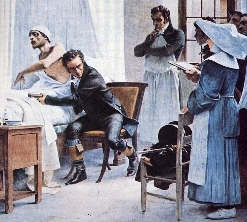

Laennec examining a tuberculous patient by "immediate" ausculatation
with the unaided ear in the Necker Hospital, Paris in the
photo of the left.
In his left hand is the stethoscope that he
used for "mediate" auscultation. Picture after the fresco by
Theobold Chartan in the Sorbonne commemorating the invention of
the stethoscope in 1816. The photo on the right shows Laennec examing a
young boy by "mediate" auscultation with his stethoscope. This
reproduction
is taken from a painting by Robert A. Thom commissioned by Parke, Davis
and Company as part of their "History of Medicine in Pictures" series,
copyrighted
in 1960.


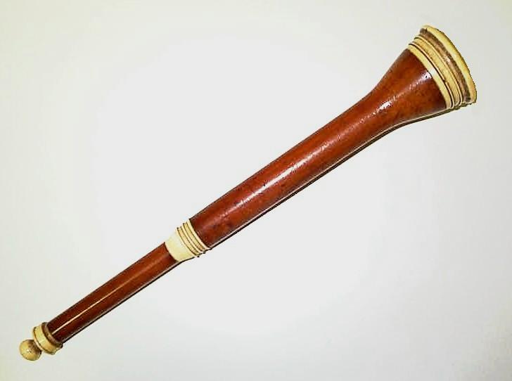
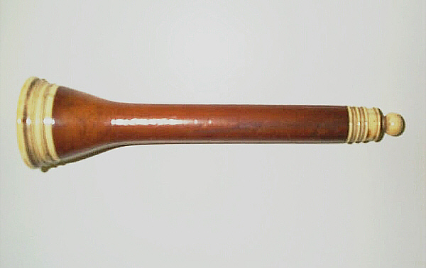
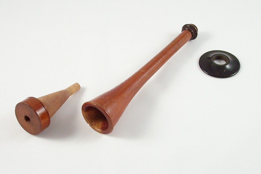
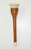
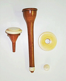
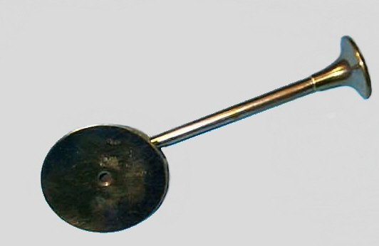
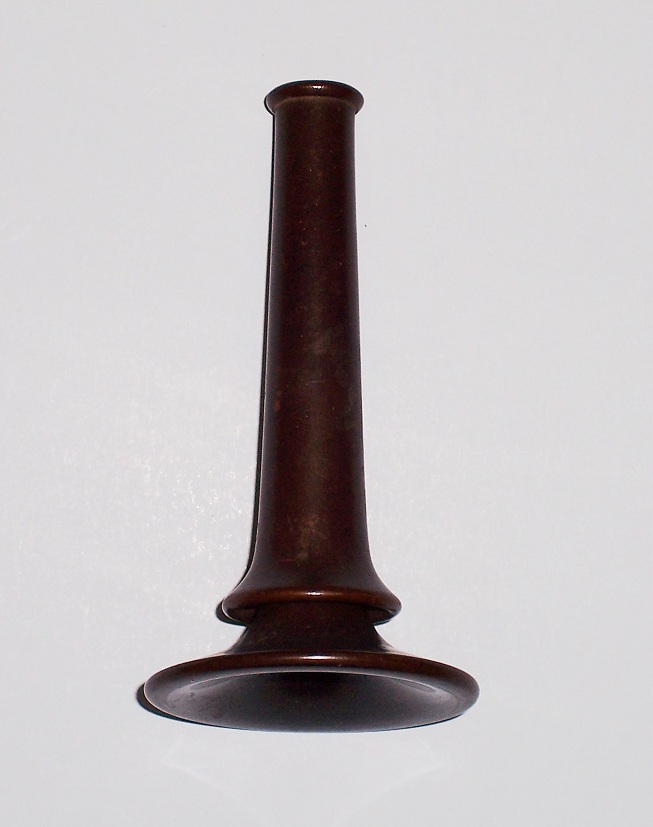
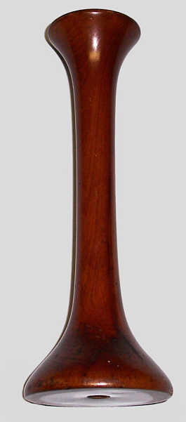
A second version Laennec
stethoscope made of a turned finely grained, light wood,
circa 1826. The cylindical stethoscpe has three parts
fitting together by rounded wood pressure fitting and brass tube
fitting and horn rings at the juncture of the three parts. It
has an overall length of 12 inches and a diameter of 1.5 inches.
This second version is illustrated in Laennec's second
edition text on auscultation published in 1826, which described the
stethoscope as having an overall length of 12 inches and a
diameter of 1.5 inches. On the left the stethoscope is assembled
for auscultation. On the right the stethoscope is taken apart
showing the rounded wood pressure fitting which holds the two parts of
the body together and the brass tube fitting which holds the chest plug
in place in the funnel shaped end of the stethoscope.


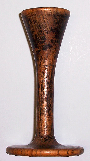
This stethoscope is the third version of the Laennec stethoscope, most likely developed in England as published in the third English edition of Laennec's text after his death (note that the Laennec design is shown on the right hand side of the illustration. The larger peices of the other stethoscope is a diagram of the original version of the Piorry stethoscope an example of which is shown below in the Piorry stethoscope section). It is marked Weiss, London, under a Crown and GR, which stands for George Rex (King George IV) who reigned from 1820-1830, thus clearly dating this stethoscope to that period. The only other known example with this mark is in the Wellcome Medical Museum, London. On the left the stethoscope is shown assembled for auscultation and in the middle taken apart. Note that a brass tube is no longer used to hold the chest plug in place and that the parts of the stethoscope are attached by a funnel shaped, wood pressure fitting. A close up of the maker's mark in shown on the right.


Unique second version Laennec stethoscope made of cedar wood with an extension piece made of cedar, ivory and horn, circa 1826. On the left is the main body of the stethoscope taken apart. In the middle the extension piece is taken apart. On the right the main body of the stethoscope is assembled and the extension piece screwed into the chest plug. The extension piece was based on the design of Nauche to allow fetal auscultation via the vaginal portion of the uterus.


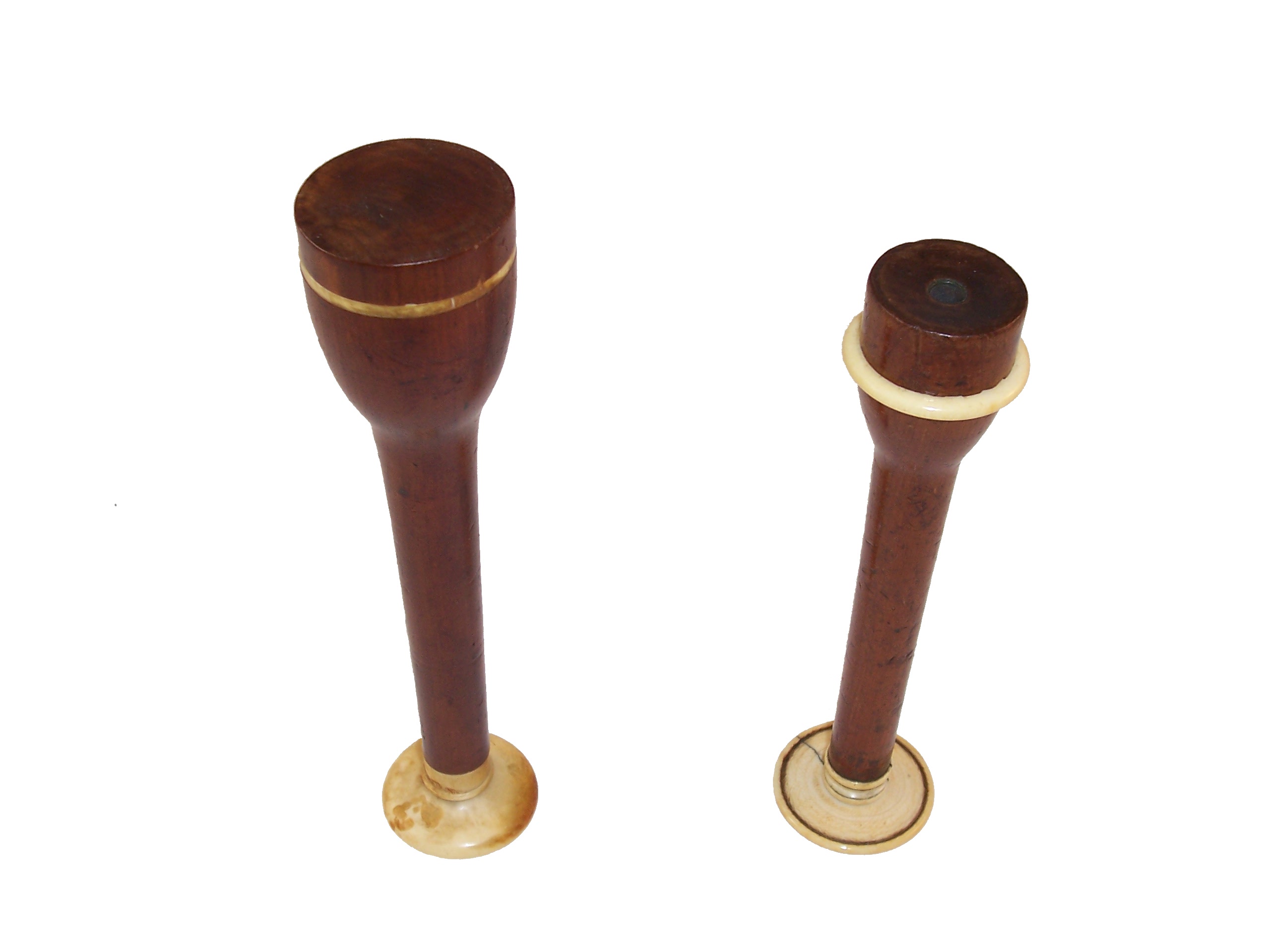
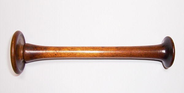
These one piece stethoscopes are
probably as far as the Laennec design of the stethoscope was
developed, circa 1830.
On the right is the model for adults and on the left is an
early obstetrical or pediatric model.
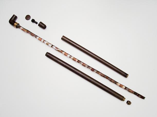
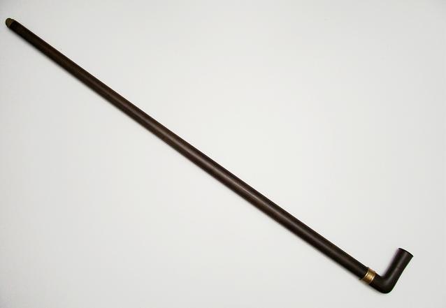

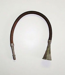
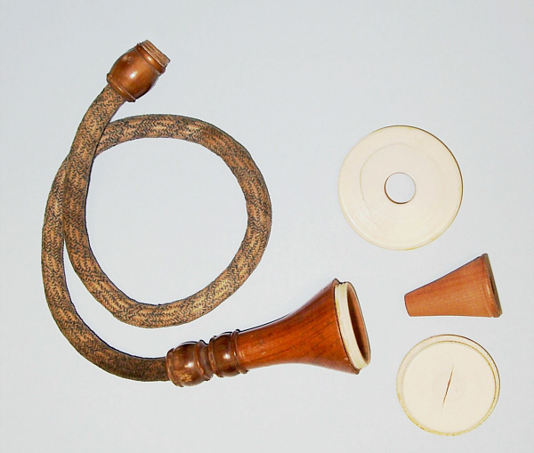
Exceedingly rare and unique medical cane made of hard rubber with removable metal assembly that holds all the original fourteen cork-stopped medicine vials. The vials have their original label and medicine content. By removing the lower tube of the cane and attaching a bell and earpiece from the handle, a seventeen inch stethoscope is assembled. The brass presentation ring just below the handle is inscribed: From Dr. Parsons to Dr. Hallock Aug. 1882. Dr. Robert Parsons is listed in the 1880 census of Salida, Colorado. Dr. Richard Sanford Hallock, also in the 1880 Salida census, was born in Orange County, New York, in 1829 and died in Salida on March 25, 1891. Dr. Hallock moved from Oakfield, Iowa, to Colorado in 1879. He served late in the Civil War as an Assistant Surgeon with the 67th U.S. Infantry of Colored Troops. The cane was clearly used by Dr. Hallock, as the brass tip is worn from walking. In the middle photo the cane is assembled for walking, with the brass tip at the top of the photo and the hard rubber handle with inscribed brass ring at the bottom. In the left photo the cane is taken apart, showing the all original medication vials it still contains. The lower tube with the brass tip is on the bottom and the middle tube and handle taken apart is at the top of the photo.In the right photo the middle tube and the earpiece and bell components of the handle are screwed together to form a monaural stethoscope.


Three portraits of Pierre Aldophe Piorry,
circa 1830s.
( Photos courtesy of the National Library of
Medicine)

Original Piorry stethoscope made of wood
and ivory, circa 1828. This is the stethoscope illustrated in Piorry's
text on percussion as shown above and published in 1828.
Shown in the middle is the stethoscope assembled with the extension
piece. The stethoscope could be used with or without the extension
piece attached.
On the left it is shown taken apart to display the main stem, extension
piece, chest plug that fits in the chest funnel end, pleximeter that
screws on to cover the chest end, ear piece that screws on the the stem
end, and finger thimble used as a plexor. On the right the same
stethoscope is shown put together for carrying (the extension piece
fits inside the main stem for carrying).


Typical Piorry Stethoscope made from cedar. On the left the stethoscope is taken apart showing the main stem, plug that inserts into the funnel shaped chest end, ivory chest piece also used as a pleximeter and ivory ear piece that screws onto the stem as the ear plate, circa 1830. On the right it is assembled for auscultation.


Cased Piorry Stethoscope made of cedar and
ivory with Percussion Hammer, circa 1835. A rare and wonderful example of
medical scrimshaw is shown on this presentation Piorry stethoscope. The
stethoscope is assembled for auscultation on the right. The scrimshaw
is shown on the left. The ivory pleximeter which screws onto the funnel
shaped chest end has a etching of a thumb lancet used for bloodletting,
poppy seed used to make morphine and Asklepios's staff showing
a rod and snake (the medical caduceus), and in latin the words
Conjurat and Amice (from your wife with love). The ivory ear plate
which screws onto the stem reveals the presentation date
May/11/1829/Paris etched on the inner surface.
On the right the assembled stethoscope and hand carved ebony percussion
hammer with cork tip are shown taken out of the case.
Later model Piorry stethoscope made from ebony. The stethoscope is
taken apart showing the ivory pleximeter with finger grasps and a
smaller ivory ear plate, circa 1840.


THE HOPE PRESENTATION STETHOSCOPE 


A presentation stethoscope given by Dr. James Hope to one of his
exceptional medical students in 1839. It is made of cherry wood and
ivory and modeled after the Piorry stethoscope. Dr. Hope designed an
ivory ear piece that was curved so as to better fit the ear. The
stethoscopes were made by James Grumbridge, a turner and stethoscope
maker in London. The silver band is engraved "Prize for
auscultation awarded to C.J. Freeman by Dr. Hope, 1839." Mr. Freeman
started his medical studies in 1837 at the Aldersgate Medical School in
London. One of his courses was the Principle and Practice of Medicine
taught by Dr. Hope. From 1838-1839, he completed 12 months of clincal
experience at nearby St. Batholomew's Hospital. During his clincal
clerkship, Mr Freeman presented a case that is discussed
in Hope's textbook on Diseases of the Heart. The patient
was in St. Bartholomew's Hospital from May 4-27, 1839. Dr. Hope
writes that "the following case is a curiosity, as it presents a
greater number of different murmurs (namely ,six, including that rare
one- the direct mitral) than I have heard in any other instance: yet it
will be seen that they were unraveled with the greatest clearness by a
student! This gentleman was James Freeman, a pupil of my class on the
practice of medicine, who brilliantly won my prize for auscultation for
the year. I give this case in his own words, the accuracy of which I
have verified by a personal examination of the patient." There were
only a total of four such presentation stethoscopes awarded and the
stethoscope pictured above is one of three known to exist today. Dr.
Hope's oil portrait by Thomas Phillips, c. 1841 is also shown.
(click here to read about Dr.
Hope and the history of this stethoscope) .
(Photo of Hope courtesy of the National Library
of Medicine)
SIR JAMES McGRIGOR'S STETHOSCOPE


Sir James McGrigor was the father of the Royal Army Medical Corps.
Dr. McGrigor introduced the stethoscope into milirtary practice in
Britain in 1821. The stethoscope is shown above both together and taken
apart, with the chest plug made entirely of wood and the ear plate made
of horn. It is an interesting variation of the Laennec and Piorry
designs, in that it has the Laennec plug that inserts into the funnel
shaped bell on the chest end (to asculatate heart sounds) and the
thinner stem and ear plate like the Piorry stethoscope. Note that
on the stem in an uneven, engraved mark that reads "McGrigor
Maker." There is no record of an instrument maker named McGrigor. The
uneven, engraved mark suggest that Dr. McGrigor designed and had this
stethoscope made for himself (or even made the stethoscope himself).
The oil portrait of McGrigor is by the early 19th century English
portraitist John Jackson presented to his family by the medical
officers of the Army.
The Piorry stethoscope became the standardfor doctors to use for
auscultation in the mid 19th century. There were manty
modifications of the Piorry design which made it easier to use and
carry the stethosopce.

An early Piorry type stethoscope with a large but simple bell chest end, thick stem and ivory ear piece, circa 1830 .

A pair of stethososcopes, circa 1830,
that combine the characteristics of the Laennec (chest plug with tube)
and Piorry (chest bell and ivory ear piece) stethoscopes.
On the left they are put together, on the right
taken apart showing the plugs. Note that the stethoscope on the left
has a solid wood plug, while the one on the right has the usual
plug with a brass tube.
Elliotson's stethoscope, circa 1835. On the left it is put together for carrying and right taken apart.

Typical European Piorry type stethoscope made of wood with a screw-on ivory earplate, circa 1875. This stethoscope was brought from Germany in the late 19th century by a pathologist from Hamburg, who immigrated and practiced in American Hospitals.
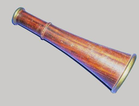
Charles James Blasius Williams developed another approach to the
design of the stethoscope. He introduced a two-piece monaural
stethoscope in 1843 with a trumpet shaped chest end that fit more
comfortably and snuggly against the chest wall. His stethoscope had a
removable ear piece.
antiquemed.com
THE WILLIAMS STETHOSCOPE


Photograph of Charles James Blasius Williams, circa 1840.
(Photo courtesy of the National Library of
Medicine)


Williams Stethoscope, circa 1845.
On the right the stethoscope has the ear-piece removed.
In the middle the ear-piece is inserted in the smaller end, leaving a
trumpet shaped end for examination of the chest.
On the left the ear-piece inserted in the trumpet shaped end, leaving
the smaller end for listening to the heart.
Flexible monaural stethoscopes were introduced around
1832. These were tubes of coiled spring covered with woven silk,
usually 14 to 18 inches long, with a chest piece at one end and usually
a very short, straight earpiece at the other. Flexible stethoscopes are
often confused withconversation tubes, which looked the same, but were
much longer than stethoscopes.
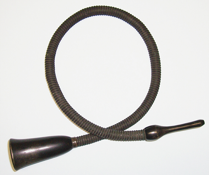
Three examples of flexible stethoscopes.
On the left is an early model made with pewter ear piece and chest
piece, circa 1832.
Golding Bird's model with wooden ear piece and chest piece is shown in
the middle, circa 1875.
Arnold's model also made with a wood ear piece and chest piece is on
the right. circa 1885.
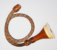
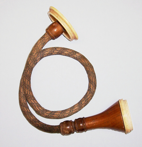
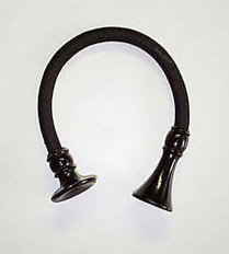
A unique Piorry Flexible stethoscope made of wood, ivory and horn,
circa 1835. Note that the typical Piorry ear plate and chest piece are
attached to the flexible tube shown in the middle photo. On the left
the stethoscope is shown assembled for carrying. On the right the
pieces are taken apart.
|
READ AN ARTICLE ON THE DIFFERENCES BETWEEN FLEXIBLE MONAURAL STETHOSCOPES AND CONVERSATION TUBES |


The stethoscope on
the left is a typical unmarked, wood Fergusson monaural, circa
1890.
In
the middle is a Fergusson stethoscope with the name T. M. Pickthall
hand engraved on the top of the ear plate, circa 1880 (click on the
image to see the engraving).
The
Fergusson shown just to the right is made by Coxeter
& Son with a hand engraving of a mascot carrying a flag and
the initials F.C.H.S. circa 1870 (click on the image to
see the engravings).
The Fergusson stethoscope to the far right is made S. Maw&
Son, circa 1870 (click on image to see mark).
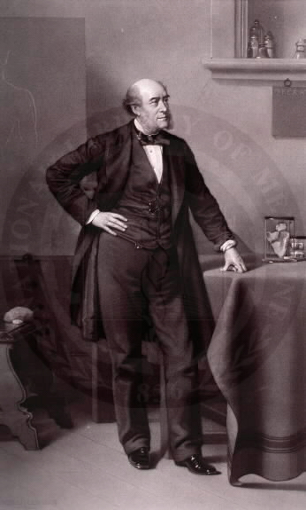
DR. W.H. HILL'S STETHOSCOPE
William Henry Hill was born on September 11, 1862 in Ormskirk, Lanchishire, England.
He entered the University of Edinburgh Medical School in 1880 at the
age of 18, and graduated in 1886 with a degree in medicine
(Bachelor in Medicine [MB]) and surgery (Master in Surgery [CM]).
During medical school he completed his medical and surgical clinical
clerkships at the Royal Infirmary of Edinburgh. The University of
Edinburgh required that a candidate for Degrees in Medicine and Surgery
must have attended for at least 3 years the medical and surgical
practice at the Royal Infirmary of Edinburgh (or a university
recognized general hospital elsewhere). After graduation, Dr. Hill
practiced in Silloth, Cumberland, England in 1887 and then in
Churchfields, Old
Basford, Nottingham, England from 1888-1915, as recorded in the annual
London and Provincial Medical Directories. He married Fanny Cox in
1892, and they had three sons, one of whom a became a doctor (Charles
Ernest Hill) also educated at the University of Edinburgh and Royal
Infirmary of Edingburgh. Dr. Hill was member of the General Council of
the University of Edingburgh, Medical Officer and Public Vaccinator for
the Basford District of the Basford Union and Medical Referee for the
British Empire, Pearl, Prudential and Victoria Assurance Companies. He
practiced medicne and
surgery for 29 years, and died on March 17, 1915 during the
"Great War."


Shown above is a Fergusson wood stethoscope, circa
1880, with hand engraved initials R.E.I. / W.H. Hill. Wards 32.
33 (click on image to see the detail of the engraving).
This stethoscope belonged to William Henry Hill, MB. CM. who
used it at the Royal Edinburgh Infirmary on General Medicine wards 32
& 33 during his clinical clerkships as a medical student at the University of
Edinburgh. He did his medical and
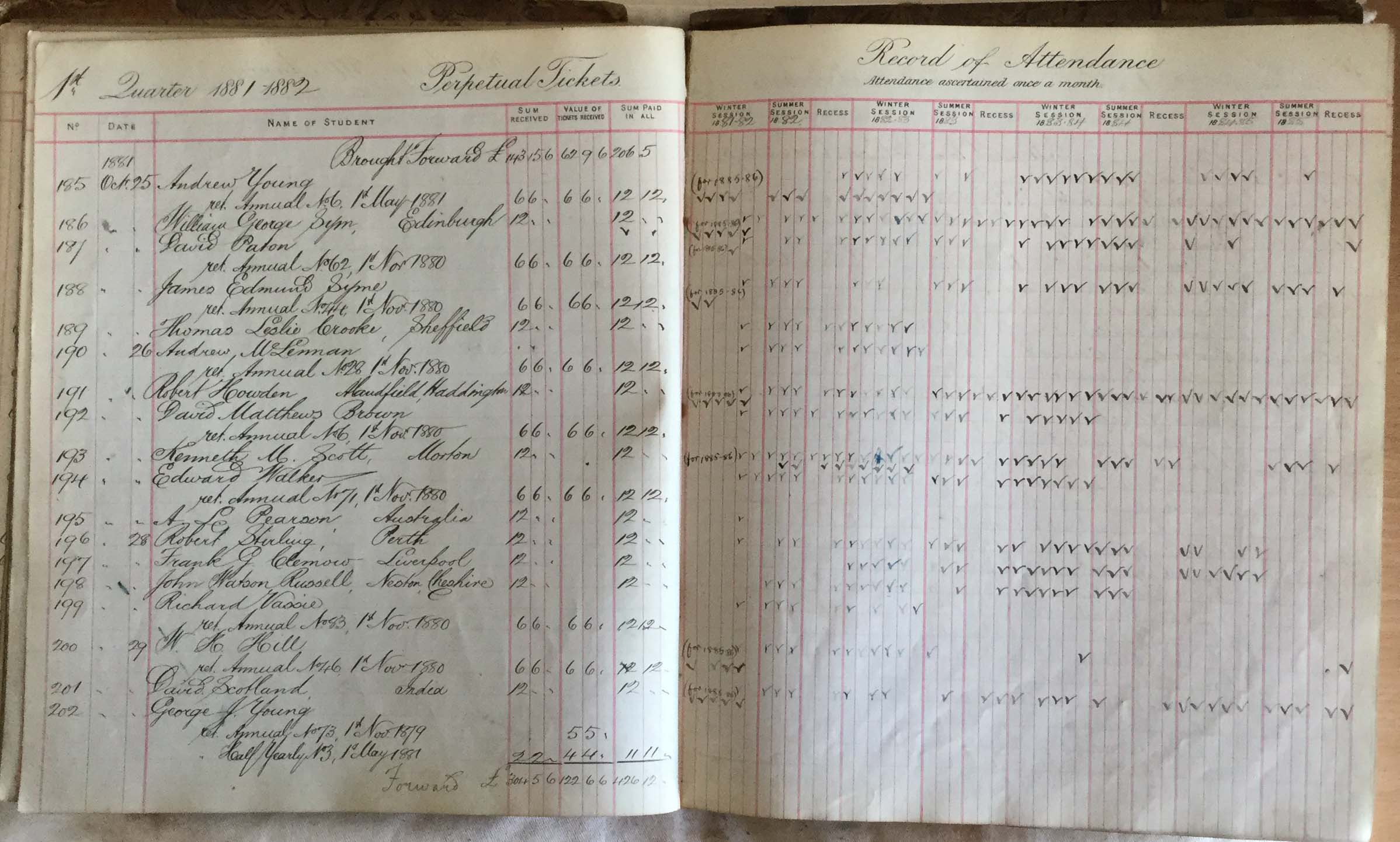
surgical clinical clerkships at the Royal Infirmary as noted in the Student Register of Tickets. These
tickets enabled medical students to participate in the "Medical
and Surgical Practice" at the hospital and "visit the Wards and
Operating Theatres, and attend Post-Mortem Examinations". W.H.
Hill purchased Royal Infirmary annual ticket No. 46 1st Nov 1880
and perpetual ticket No. 200 21st Oct 1881, as recorded in
the Royal Infirmary ledger of Perpetial Tickets 1881-1882 (click
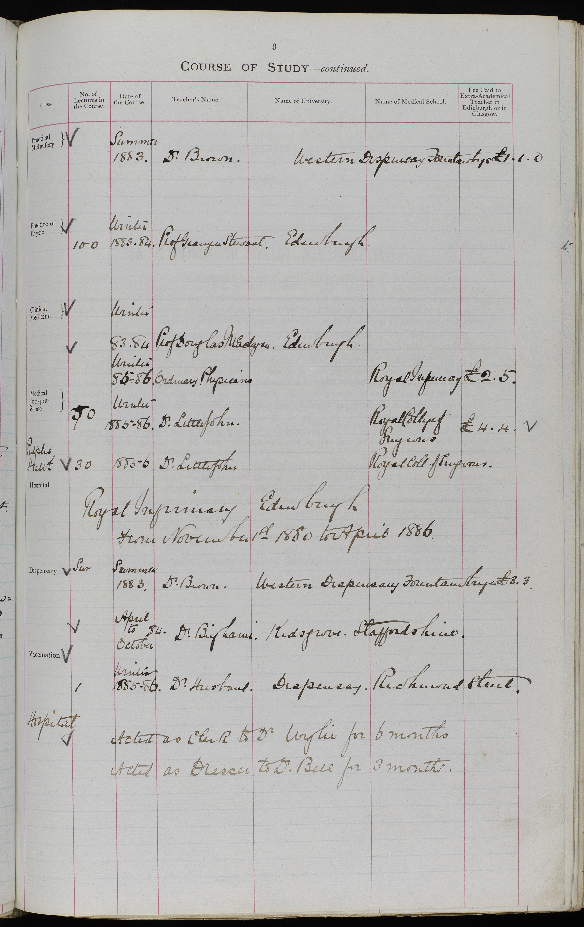
on image to see the ledger detail). Also
shown are three pages of the Undiversity of Edinburgh Medical
Graduation Records for Dr. Hill, revealing the courses he completed
from 1880-1886 and Professors who taught these courses (click on image
to see the detail of the courses). In the middle of page 3 is a
notation "Royal Infirmatry Edinburgh from November 1st 1880 to April
1886." Also at the bottom of page 3 under the handwritten title
"Hospital" is a notation that he acted as the
Clerk to Dr. John Wyllie for 6 months and as Dresser to Dr. Joseph Bell
for 3 months. Dr. Wyllie would later become the Chair of Medicine at
the University of Edinburgh Medical School and
Dr. Bell's medical student Arthur Conan Dolye said he modeled the
chracter Sherlock Holmes after Dr. Bell because he was a great observer
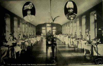
and diagnostician. (Images of the Register of Tickets and Courses of Study were provided by the Edinburgh University Library Special Collections). On the upper left is a postcard of the Queen Mary Ward at the Royal Infirmary
in 1911, looking much as it did when Dr. Hill used his stethoscope
on wards 32 & 33. King
George V and Queen Mary visited
the Royal Infirmary on July 19, 1911 following their Coronation in
London on June 22, 1911. They visited surgical ward 7 and medical
ward 30, which were then named the King George V Ward and Queen Mary
Ward, respectively, in commenoration of the Royal Family visit.
Dr.
Hill died on March 17, 1915 during the "Great War" (World War I), which is
memorialized in one of the eight Basford St. Leodegarius church bells
reading "In Memory Of William Henry Hill M.B.C.M. Who Died March 17TH
1915 During The Great War." Church
parishioners also palced a large stone plaque on its south aisle wall
which is shown below:
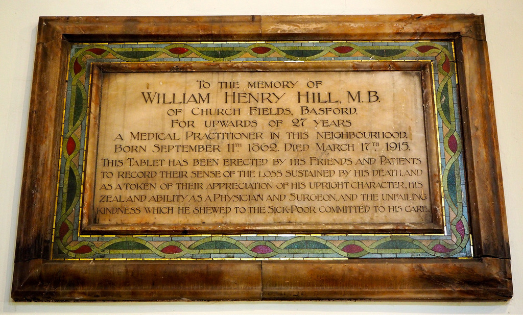
(To the Memory of William Henry Hill M.B. Church Fields, Basford. For upwards of 27 Years. A medical practitioner in this neighbourhood Born Sep.11 1862. Died March 17th 1915. This tablet has been erected by his Friends and Patients To record their sense of the loss sustained by his death and as a token of their appreciation of his upright character, his zeal and ability as a physician and surgeon and the unfailing kindness which he shewed to the Sick Poor committed to his care)
In 1729, a four bed hospital was established with funds from the Royal College of Physicians of Edinburgh at the head of Robertson's Close in the heart of city. It was known as the Hospital for the Sick Poor, Physicians' Hospital or Little House. It was the first volunatry hospital in Scotland. Granted a Royal Charter in 1736, the Royal Infrimary of Edinburgh moved to new premises on what is now known as Infirmary Street in a 228 bed facility designed by William Adam in 1741. It was in this Royal Infirmary that Dr. James Hope first used the stethoscope and learned the art of auscultation at the bedside of patients, when he served as a House Physician and Surgeon from 1824-1825. In 1872, David Bryce was commissioned to design a new hospital, and in 1879 the Royal Infirmary moved to a "clean air" site at Lauriston Place. The main building of the Royal Infirmary at Lauriston conformed to the Florence Nightingale pavilion design of medical and surgical wards. The Infirmary set apart a portion of the beds for clinical instruction by Professors of the University of Edinburgh and its Medical Department gave special instruction in Physical Diagnosis. Postmortem examinations were conducted by the patholgist in the Anatomical Theatre. Separate Wards were devoted to certain types of illness. Wards 32 (men) and 33 (women) were designated for General Medicine and ultimately in 1963 for Medicine of the Ederly (MOE). In 2002, the Royal Infirmary of Edinburgh moved to its current home at Little France in the southern suburbs of Edinburgh. The MOE service still exists today in wards 201 and 202 in the new Royal Infirmary. The University of Edinbuirgh College of Medicine required that a medical student attend at least three years of medical and surgical practice at a General Hospital. Continuing the long standing relationship of the Royal Infirmary and Medical School since 1750 , the University of Edinburgh also moved the Medical School to Little France and located its new home, The Chancellor Building, adjacent to the Infirmary.

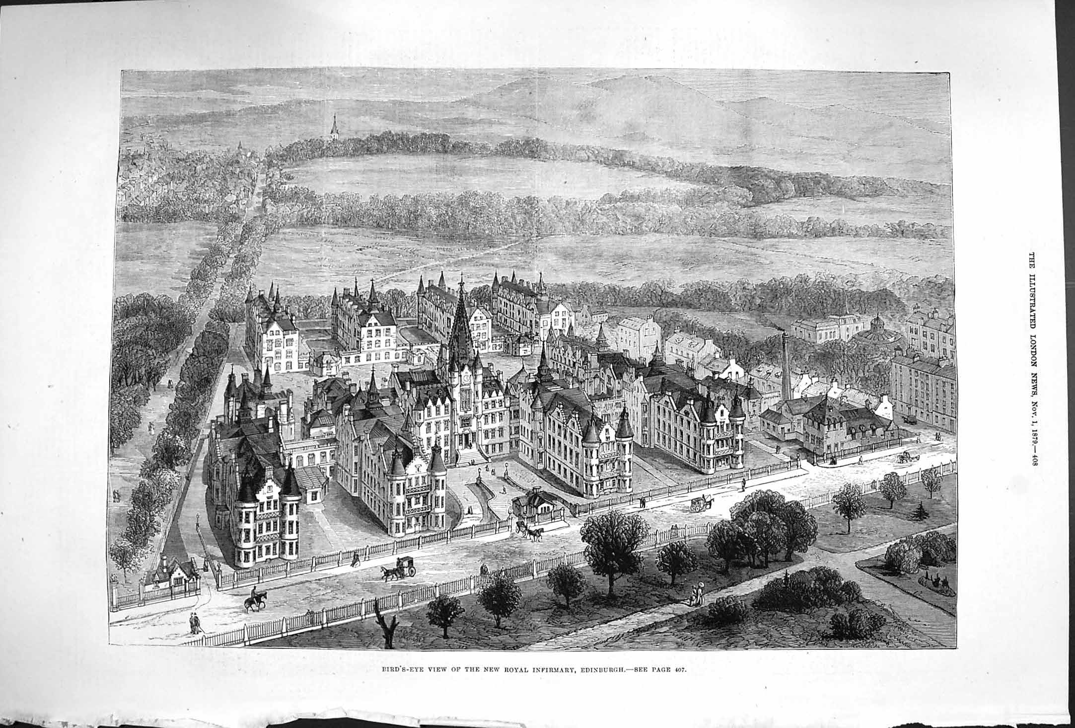

Shown above in the middle is an
engraving of the Royal Infirmary of Edinburgh at Lauriston Place from
the Illustrated London News in 1879, the year the Royal Infirmary at Lauriston Place opened.. The four wings in
the front of the Royal Infirmary are part of the surgical hospital and
the four wings in the back are part of the medical hospital. The
last wing on the right in the medical hospital contained wards 31,
32 and 33 (first, second and third floors, respectively). On
the the left is a 2015 photo of the current Royal
Infirmary medical buildings that were rennovatd to become a residential
part of the
Edinburgh Quatermile development. The buiding in the foreground is the
rennovated medical wards 31, 32 and 33 (top three floors). Click on the
image and enlarge the photo to see examples of the upper part of each
window opened, which was the Florence Nightingale design to allow cross
ventilation of the Infirmary wards. The image on the right is the
architectural drawing of the "Edinburgh Futures
Institute" of the University of Edinburgh. In 2017, The Universtity
began restore the historic former Royal Infirmary surgical hospital to
create a multidisciplinary building to form a wide range of teaching
and learning spaces combining sciences, humanities, arts, business and
external partners. The Institute is scheduled to open in 2021 in the
heart of the Quartemile development.
In general, the Piorry design was the most commonly employed form.
However, there were many variations in the shape and form of
different models. Some stethoscopes were designed for special purposes.
And some were made from special materials that generally signified a
physician with "upper class" patients.
Monaural stethoscopes from 1850 to 1900 are shown below.


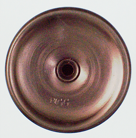

Shown above are a group of stehtoscopes made of unusual materials. An exquisite stethoscope carved from one piece of ivory,
circa 1850. It may have been a show stethoscope. A beautiful
silver plated stethoscope, circa 1860. The hollow ear piece is
constucted much like the base of similar silver items and served as a
chamber to enhance the ascultated sounds. A well crafted monaural
stethoscope made of ebony and marked "BIGG" ( click on image to see the
maker's mark ) on the concave underside of the ear piece as shown on
the right, circa 1850. This company was only in business until 1859. The unusual stethoscope on the far right is made of brass with cedar wood
integrated into the brass ear piece, circa 1890. Note the curved shape
of the wood ear piece with a gentle protrusion so that the ear sealed
tightly against the stethoscope for enhanced sound transmission during
ausculatation. The stethoscope could be taken apart and one piece screwed on top of the other for easy carrying (click on image).

Burrows stethoscope with original rubber
percussion ring around the ear plate, circa 1860. Stokes
stethoscope with original rubber percussion ring around the ear plate,
circa1880.


Interesting examples of monaural
stethoscopes that have a narrow oval chest end which was intended to
examine the chest in between the ribs so as to better auscultate the
lungs. On the left is a completely solid model, circa 1850. On the
right is a similar version, but with the central hole bored throught
the stethoscope, circa 1840. Note the middle photo which shows the
chest ends of the stethoscopes with and without the hole.


Traube's stethoscope in a case with precussion
hammer and pleximeter by H. Hauptner, Berlin, circa 1876. The Traube
stethoscope, Metallstiel percussion hammmer, and ivory pleximeter are
shoun out of the case on the left.

Hecker's stethoscope made of wood with a horn chest piece
and a horn extension to attach a flexible tube with a horn earpiece for
student teaching.
CIVIL WAR SURGEON ROBINSON STORY
There has been considerable debate about whether stethoscopes
were used by civil war doctors. The evidence that civil war
surgeons did not use stethoscopes is usually referenced to
the fact that the Harvard Medical School catalogue did not list
ownership of a stethoscope until after the civil war in 1869. However,
medical students owned their own stethoscopes dating back to the 1840s.
There is ample evidence in civil war army medical documents that
physical auscultatory signs related to diseases of the heart and
lungs could only have been heard with use of the stethoscope. In
his highly regarded Manual of Military Surgery published in May of
1861, Samuel Gross said that "Organic cardiac disease could easily be
detected with the stethoscope." Both
union and confederate army medical department regulations show
stethoscopes as part of the medical supplies for civil war hospitals.
In 1865, a hand written inventory from ward H at the Conesus
Centre Army Hospital, N.Y. lists a stethoscope as part of their
medical supplies. The 1863 Manual of Instructions for Military Surgeons
by John Ordronaux, M.D. lists a stethoscope as part of Instruments for
Special Diagnosis. A note in the section on Diseases of
the Chest and Back states that "It is a good plan, in auscultating
a party, to place him with his back against a wooden door or patition.
The greater resonance of the pectoral sounds obtained by this process,
will suprise those who have never bfore availed themselves of this
simple acoustic medium." The manual also contains an
illustrated Auscultatory Percusssion Chart. This information confirms
that the stethosocpe was part of civil war medical supplies.
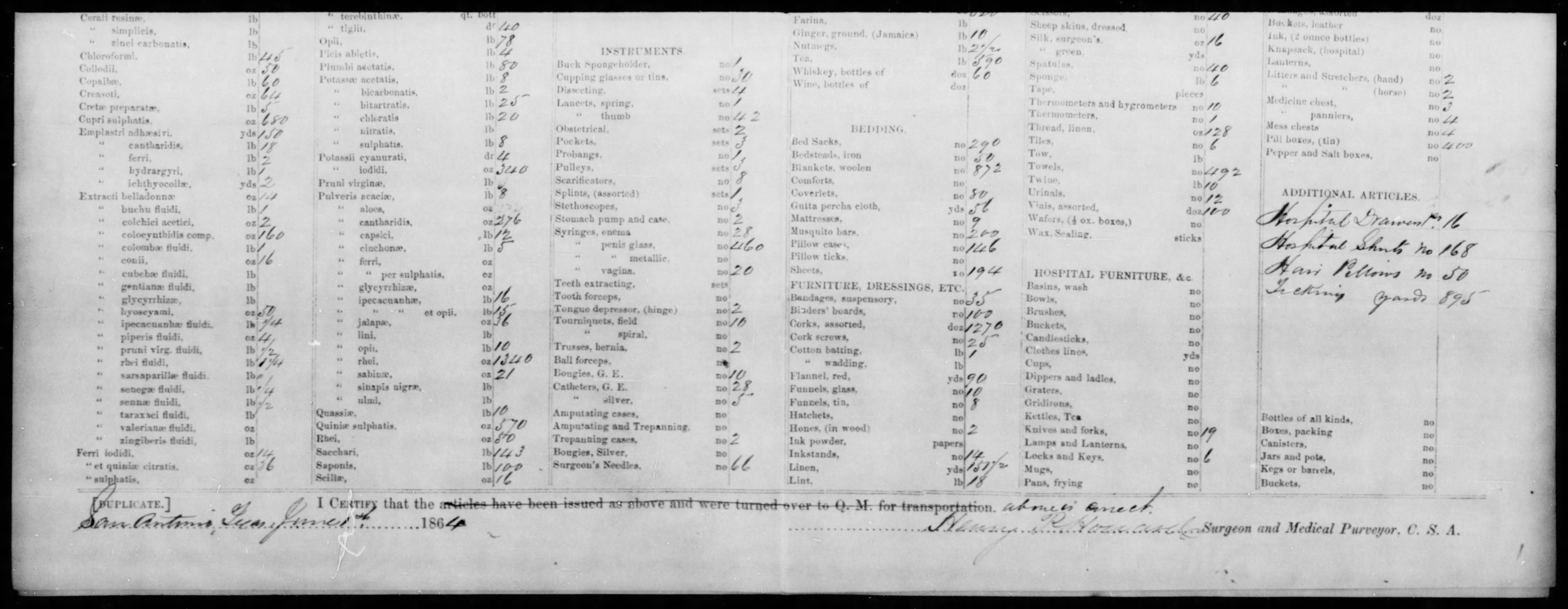
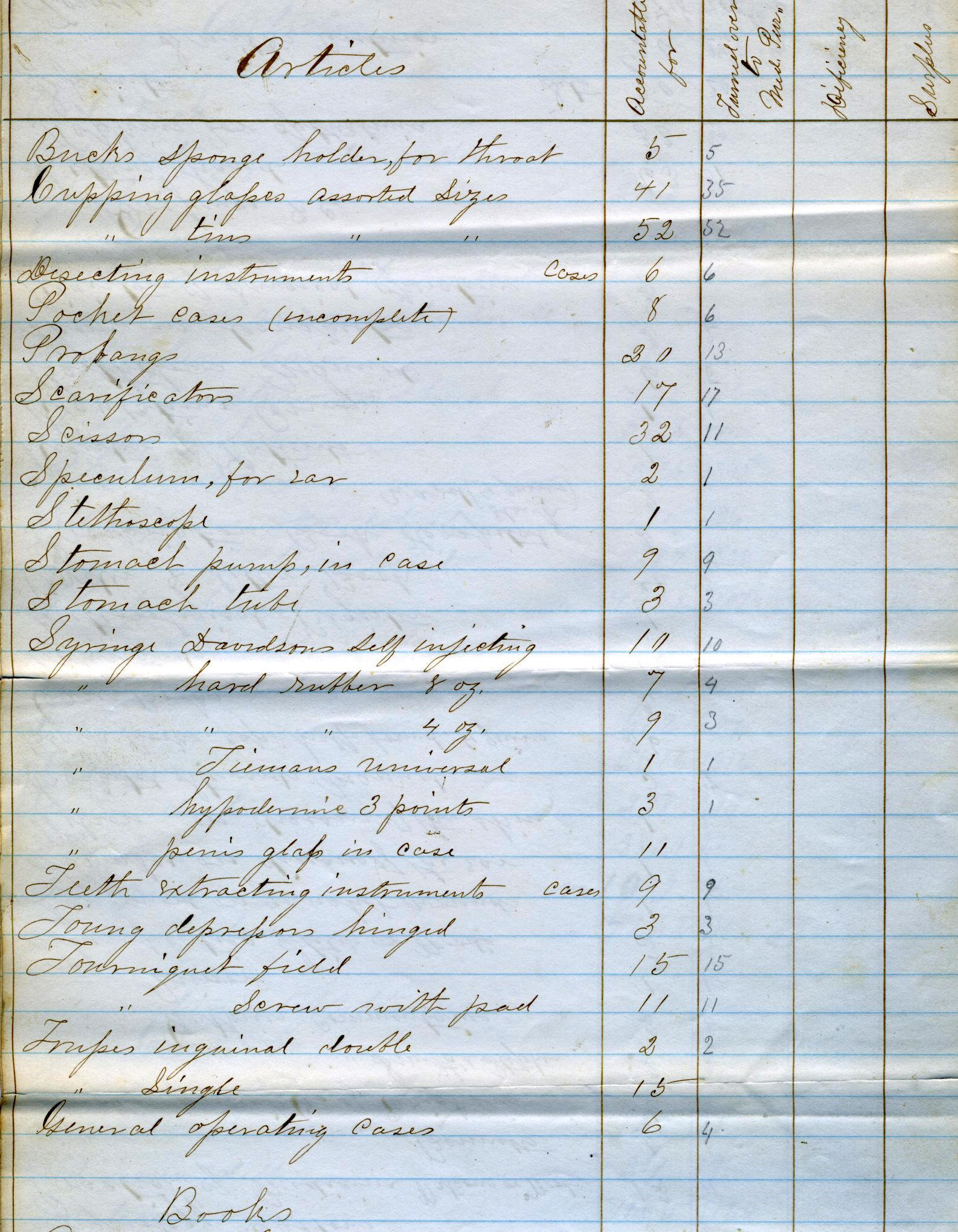
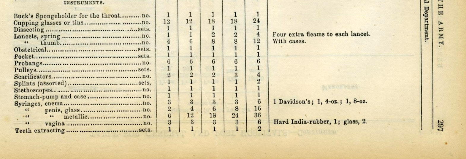
Shown above on the right is a page from
the The Army Medical Department book that lists a stethoscope along
with other instruments as part of the supplies for the civil war
hospital. On the left is a Medical Purveyor report for the Confederate
States Army that lists stethoscopes as part of hospital supplies. Also
displayed is a hand written inventory from Hospital Ward
H, Conesus, NY, 1865 showing a stethoscope as part of the ward
Articles.
(center photo courtesy of Michael Echols)
Civil war confederate surgeon William R. Robinson received
his undergraduate education at the West Point Military Academy from
1845-1849 and medical education at the Medical Department of the
Univerity of New York from 1853-1857. Dr. Robinson would have
been exposed to the use of the stethoscope for auscultation during
his clinical courses at Bellevue Hospital under the tutelage of renown
professors such as Valentine Mott, the "father" of American surgery,
and John Metcalfe, who taught the course on "Physical Diagnosis and
Diseases of the Chest." After graduaton, Dr. Robinson was
appointed Assistant Physician at the Seamen's Retreat Hospital on
Staten Island, NY based on a letter of recommendation from
his teacher Valentine Mott. In 1860, he moved to Galveston, Texas
where he was again recommended as a superb physician by Dr. Mott. At
the start of the civil war, he joined the Texas Rangers and was
appointed an Assiatant Surgeon of the Texas Volunteer Forces,
Provincial Army on Decemeber 10, 1861. He served in the 3rd
Regimen of the Arizona Brigade in northern Texas and as Director of the
confederate general hospital in Columbus, Texas. Towards the end of the
war, he was an Acting (contract) Assistant Surgeon for the union army
prison on Ship's Island, Mississippi. Dr. Robinson eventually returned
to Newark, New Jersey where he practiced until his death in 1889. Dr.
Robinson's journey is just one example of a West Point cadet
serving in the confederate army during the civil war that divided
family and friends into north and south camps.


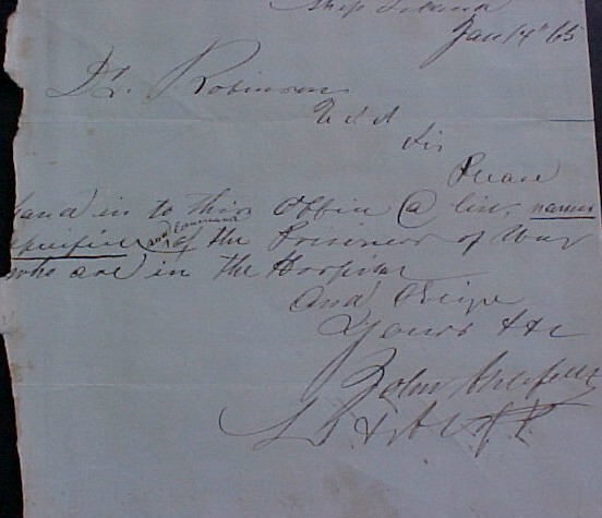


On the left are three hand written, civil war Dr.
Robinson letters. The first letter on April 28,
1861 is written by Dr. Robinson to his father from Port Sullivan,
Texas. Dr. Robinson
states "War has commenced! Abe Lincoln has thrown the first stone! The
South will fight to the last - The result will be a long and bloody
war." Another letter on 17 Nov 1862 is addressed
to "Doctor Robinson Principle Director Hospital Columbus, Texas"
and signed "Henry L. Webb Inspector Genl. Dept. of
Texas." In this letter Henry Webb tells Dr. Robinson
that "W.E.B. Howe of Col. Elmore's regiment is ordered to
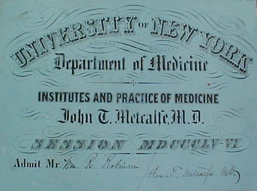
On the right are two University of the City of New York
medical school tickets for William R. Robinson's courses in the
"Practice of Medicine" 1855-56 taught and signed by Dr. Metcalfe and
"Operative Surgery" 1855-56 taught and signed by Dr. Valentine
Mott, the "father" of American surgery (note the hand holding a scapel
in the design at the top of the ticket). On the far right is a letter
that Dr. Mott wrote on April 18, 1857 recommending Dr. Robinson
for his first position after medical school at the Seamen's Retreat
Hospital on Staten Island.


A Roberts stethoscope made of ivory, circa
1880. On the left the stem is shown inserted thru the earpiece for easy
portability and on the right the stem is screwed upright into the ear
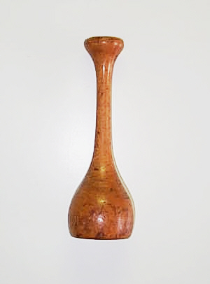
piece for auscultation.The left wooden model is known as the Dutch
Stethoscope, because of the tulip shaped bell, circa 1890. This
stethoscope came apart in three pieces for carrying. Brass monaural
stethoscope with a swivel joint for portability, circa 1890. On
the left the stethoscope is upright and on the right the swivel joint
is bent at a right angle for carrying.

The gutta-percha model on the right is a simple
stethoscope, with an ear plug end, circa 1880. Unusual
wood stethoscope with a large bell ear piece, circa 1850
the typical chest end is at the top of the photo. Cedar
stethoscope with curved, protruding ear piece made of gutta percha,
which was intended to create a better fit in the ear, circa 1860. An
interesting stethoscope with a brass rim at the ear-end and a brass
lined funnel shaped chest-end. A hand carved, funnel shaped
stethoscope made from a unique vermont wood.


On the left is a Quain's telescoping
stethoscope where the chest-end screwed onto the stem ear-end for
ausculatation and could be unscrewed for ease of carrying, circa 1880.
The photos show the two parts screwed together for auscultation
and the chest-end screwed on top of the stem ear-end for carrying. The
two photos on the right show another version of a portable stethoscope
with the parts screwed together and taken apart. The two stem
pieces fit into the two holes in the ear piece for easy
carrying.


A ninetenth century photograph of Dr. William Lennard
holding an unusually long monaural stethoscope in his right hand.
This type of long stethoscope was intended to keep the doctor a
distance from the infested patient.
(Photo courtesy of the Wellcome Library)
A very long (15 inches) pauper's stethoscope,
circa 1850 is shown to the left of the photo. The stethoscope
unscrewed in the middle so that it could be carried more easily, much
like the original Laennec stethoscope. On
the right, is a very long (14 inches) stethoscope circa 1880.
The lower part of the stem has a hand carved letter A, which
is similar to the branding of animals on a ranch. This extra
long stethoscope is most likely a vetenary stethoscope. To the right is
a long (10 inches) stethoscope made of gutta percha which was used to
examine patients with fever, circa 1890. It is marked Maw on the ear
piece.
Stethoscopes were also developed for obstetrical and pediatric
auscultation. Laennec's friend Jacques-Alexandre Lejumeau de
Kergaradec was the first doctor to use the stethoscope for fetal
auscultation and this technique was discussed by Laennec in his second
edtion text on ausculataion. The fetal stethoscopes that emerged
usually had a very wide or flaring bell and a wide earplate, which
prevented the stethoscope from rocking on the abdomen of the mother
during fetal auscultation. Stethoscopes for children tended to be
shorter than those for adults and were probably used as either
pediatric or obstetrical stethoscopes.


A very short model (4 inches) with funnel shaped end and wide earplate called the Pajot Stethoscope used for fetal auscualtation, circa 1880. Pajot designed a shorter stethoscope than DePaul to avoid rocking on the mother's abdomen during fetal auscultation. An example of Pinard's aluminum fetal stethoscope (6 inches), with the characteristic very wide, deep bell circa 1900. The Pinard stethoscope rapidly became the fetal stethoscope of choice becqause the widely faring bell prevented rocking on the mother's abdomen during auscultation. DePaul stethoscope (5 inches) used for fetal auscultation, circa 1885. DePaul designed the stethoscope with a wider than usual bell to avoid it rocking on the mother's abdomen during fetal auscultation.


A short (5 inches) ebony stethoscope
with a small ivory earpiece most likey used for pediatric or
obstetrical auscultation, circa 1840. Another short ebony
stethoscpe (4.75 inches) with a silver lined chestpiece most likely
used for pediatric or obstetrical auscultation, circa 1850. Solid
silver stethoscope that is very short (3.5 inches), most likely
used for obstetrical or pediatric auscultation.

This stethoscope belonged to Dr. Gustav Lowenstein from
Frankfurt, Germany. Dr. Lowenstein fled Germany to Austria in 1933 and
then emigrated with his family to America in 1935. The stethoscope was
obtained in 2006 from his 78 year old physician son who said that the
stethoscope originally belonged to his grandfather, who was also a
physician. This cedar wood stethoscope is a Hosford's type, with a
large bell ear piece designed to cover the ear to exclude external
sounds, circa 1900.
The monaural instrument was used exclusively for about 30
years, and were used into the late 19th and early 20th centuries. In
fact, they are still used today in countries such as those of the
Former Soviet Union, and are still being used by midwives in the
United Kingdom and Europe. However, eventually physicians decided
to find out if an instrument using both ears would be better than the
simple monaural.
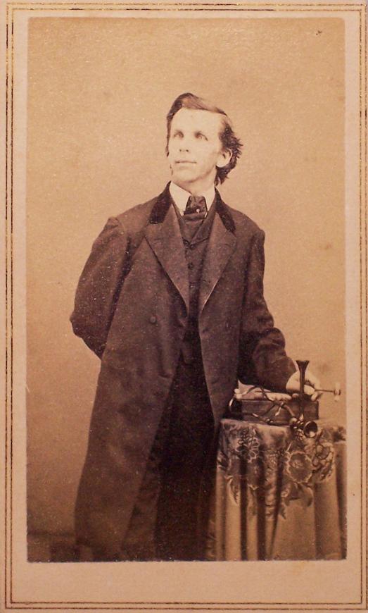
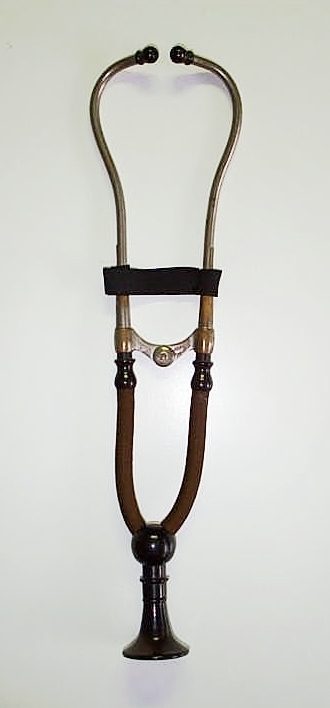

Carte-de-Viste photo of a physician
posing with a Hughes monaural and Cammann binaural stethoscope as well
as a Dejeurne percussion hammer, circa 1865. As shown on the backmark,
the photo was taken by the well known 19th century portrait
photographer Abraham Bogardus, whose studio was located at 363 Broadway
in New York from 1862 to 1869. Also shown are examples of
these instruments from the same period. On the left is a Hughes
monaural stethoscope, on the right a Cammann binaural stethoscop
and on the far right a Dejeurne percsussion hammer.
We are always interested in acquiring new items for the collection and welcome information about items for sale. If you have any comments or questions, please do not hesitate to contact us.
|
CONTACT US |